. 2022 Jun 9;5(1):564.
doi: 10.1038/s42003-022-03522-6.

Stabilization of CDK6 by ribosomal protein uS7, a target protein of the natural product fucoxanthinol

Affiliations

Stabilization of CDK6 by ribosomal protein uS7, a target protein of the natural product fucoxanthinol

Yosuke Iizumi et al. Commun Biol. .

Abstract

Cyclins and cyclin-dependent kinases (CDKs) regulate the cell cycle, which is important for cell proliferation and development. Cyclins bind to and activate CDKs, which then drive the cell cycle. The expression of cyclins periodically changes throughout the cell cycle, while that of CDKs remains constant. To elucidate the mechanisms underlying the constant expression of CDKs, we search for compounds that alter their expression and discover that the natural product fucoxanthinol downregulates CDK2, 4, and 6 expression. We then develop a method to immobilize a compound with a hydroxyl group onto FG beads® and identify human ribosomal protein uS7 (also known as ribosomal protein S5) as the major fucoxanthinol-binding protein using the beads and mass spectrometry. The knockdown of uS7 induces G1 cell cycle arrest with the downregulation of CDK6 in colon cancer cells. CDK6, but not CDK2 or CDK4, is degraded by the depletion of uS7, and we furthermore find that uS7 directly binds to CDK6. Fucoxanthinol decreases uS7 at the protein level in colon cancer cells. By identifying the binding proteins of a natural product, the present study reveals that ribosomal protein uS7 may contribute to the constant expression of CDK6 via a direct interaction.


Conflict of interest statement

The authors declare no competing interests.

Figures
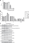
Fig. 1
Fig. 1. Fucoxanthinol induces G1 cell cycle arrest with the downregulation of CDK2, 4, and 6 protein expression.
a Human colon cancer HT-29 cells were treated with the indicated doses of fucoxanthinol for 72 h. The proliferation of cells was measured using the CCK-8 assay. b HT-29 cells were treated with fucoxanthinol for 24 h, and the cell cycle was analyzed by flow cytometry. c A western blot analysis of HT-29 cells treated with fucoxanthinol for 24 h. The signal of each western blot was quantified using ImageJ software (Version 1.52a) and normalized by the value of β-actin. The value of each signal was indicated below the blot. NT: non-treated, DM: 0.1% DMSO, Data are means ± S.D. (n = 3 biologically independent experiments). *P < 0.05 significantly different from NT.
Fig. 2
Fig. 2. Ribosomal protein uS7 is the major binding protein of fucoxanthinol.
a The scheme for the immobilization of fucoxanthinol onto FG beads® with carboxyl groups. b Whole-cell extracts of HT-29 cells were incubated with empty (−) or fucoxanthinol-fixed (+) FG beads®, and the binding proteins of fucoxanthinol were purified and detected by silver staining. c The major binding protein indicated by an arrowhead in b was analyzed by mass spectrometry, and the molecular masses of the peptides detected are shown. d Whole-cell extracts of HT-29 cells were incubated with fucoxanthinol-fixed beads, and the binding of uS7 was detected by western blotting. e The recombinant His-tagged uS7 protein (His-uS7) was incubated with fucoxanthinol-fixed beads, and the binding of uS7 was detected by western blotting. HOSu: N-hydroxysuccinimide, EDC-HCl: 1-ethyl-3-(3-dimethylaminopropyl)-carbodiimide hydrochloride, DMAP: 4-dimethylaminopyridine.
Fig. 3
Fig. 3. The molecular binding model for the interaction between uS7 and fucoxanthinol.
a The top-ranked pose in the docking simulation. Fucoxanthinol (green stick) and uS7 (gray cartoon) are shown. b Hydrophobicity around the pocket to which fucoxanthinol binds. Brown and blue colors correspond to the hydrophobic and hydrophilic regions of uS7, respectively. Images (a, b) were made using BIOVIA Discovery Studio 2018 software (Dassault Systèmes, Vélizy-Villacoublay, France).
Fig. 4
Fig. 4. The knockdown of uS7 reduces the CDK6 protein in colon cancer cell lines.
ad HT-29 cells were transfected with negative control siRNA (siCtrl) or two different siRNAs targeting uS7 (siuS7 #1 and #2). After 48 h, the depletion of uS7 was confirmed (a). After 72 h, cell proliferation was examined using the CCK-8 assay (b). After 48 h, the cell cycle was analyzed by flow cytometry (c), and protein levels were investigated by western blotting (d). e SW480 cells were transfected with each siRNA. After 48 h, cells were lysed and subjected to western blotting. In d, e, the signal of each western blot was quantified using ImageJ software and normalized by the value of β-actin. The value of each signal was indicated below the blot. Data are means ± S.D. (n = 3 biologically independent experiments). *P < 0.05 significantly different from siCtrl.
Fig. 5
Fig. 5. uS7 directly binds to CDK6 and is involved in the stability of the CDK6 protein.
a HT-29 cells were transfected with siCtrl or siuS7 #2. After 18 h, cells were treated with 20 μg/ml cycloheximide and lysed at the indicated times. The expression levels of CDK2, 4, and 6 were analyzed by western blotting. The sample, designated as “0 h half”, is identical to half the amount of the sample at 0 h. b The expression levels of CDK2, 4, and 6 at each time point were quantified. The expression level at 0 h was defined as 100%. c HT-29 cells were transfected with siCtrl, siuS7 #1, or siuS7 #2. After 24 h, cells were incubated with 10 μM MG-132 for 24 h, lysed, and subjected to western blotting. The signal of each western blot was quantified using ImageJ software and normalized by the value of β-actin. The value of each signal was indicated below the blot. d HT-29 cells were lysed, and the endogenous immunoprecipitation assay was performed with anti-CDK2, CDK4, CDK6, cyclin D, and cyclin E antibodies, respectively. Ribosomal proteins uS7 and uS9 in immunoprecipitates were detected by western blotting. e A GST pull-down assay was performed by incubating the recombinant Myc-DDK-tagged CDK6 protein with GST or GST-fused uS7 (GST-uS7)-bound beads. The CDK6 protein that bound to these beads was detected by western blotting.
Fig. 6
Fig. 6. Fucoxanthinol decreases the uS7 protein, resulting in G1 cell cycle arrest.
HT-29 cells were treated with the indicated concentrations of fucoxanthinol for 6 h, and the protein (a) and mRNA (b) levels of uS7 were examined by western blotting and qRT-PCR analyses. c HT-29 cells were transfected with siCtrl or siuS7 #2. After 48 h, cells were incubated with 10 μM fucoxanthinol for 24 h, and the cell cycle was analyzed by flow cytometry. NT: non-treated, DM: 0.1% DMSO, Data are means ± S.D. (n = 3 biologically independent experiments). *P < 0.05 significantly different from NT.

References

    1. Massague J. G1 cell-cycle control and cancer. Nature. 2004;432:298–306. doi: 10.1038/nature03094. - DOI - PubMed
    1. Sherr CJ, McCormick F. The RB and p53 pathways in cancer. Cancer Cell. 2002;2:103–112. doi: 10.1016/S1535-6108(02)00102-2. - DOI - PubMed
    1. Nakayama KI, Nakayama K. Ubiquitin ligases: cell-cycle control and cancer. Nat. Rev. Cancer. 2006;6:369–381. doi: 10.1038/nrc1881. - DOI - PubMed
    1. Vermeulen K, Van Bockstaele DR, Berneman ZN. The cell cycle: a review of regulation, deregulation and therapeutic targets in cancer. Cell Prolif. 2003;36:131–149. doi: 10.1046/j.1365-2184.2003.00266.x. - DOI - PMC - PubMed
    1. Stepanova L, Leng X, Parker SB, Harper JW. Mammalian p50Cdc37 is a protein kinase-targeting subunit of Hsp90 that binds and stabilizes Cdk4. Genes Dev. 1996;10:1491–1502. doi: 10.1101/gad.10.12.1491. - DOI - PubMed

Publication types

MeSH terms

LinkOut - more resources