Review
. 2022 Nov;23(11):651-664.
doi: 10.1038/s41576-022-00501-9. Epub 2022 Jun 9.

tRNA dysregulation and disease

Affiliations
Review

tRNA dysregulation and disease

Esteban A Orellana et al. Nat Rev Genet. 2022 Nov.

Abstract

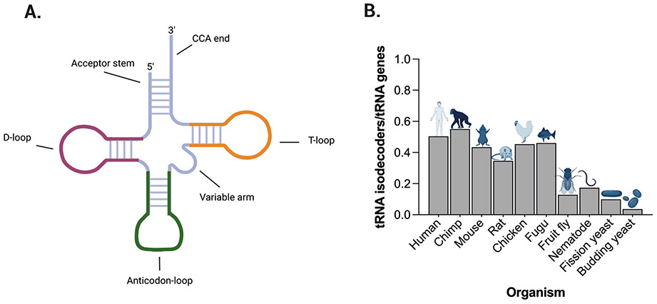
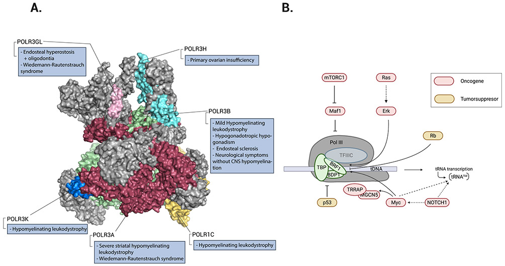
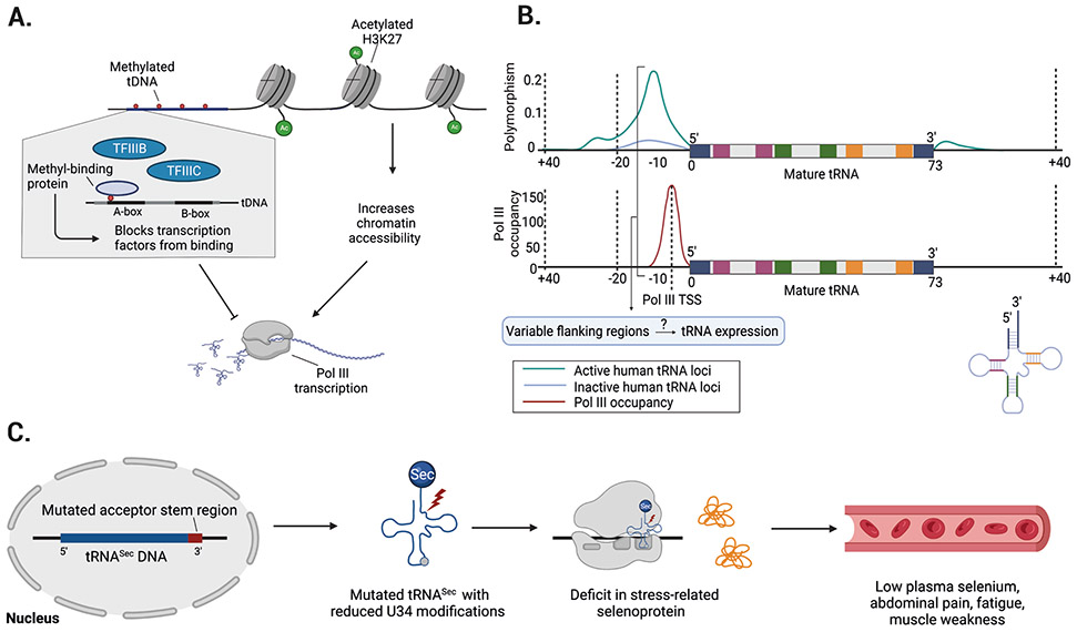
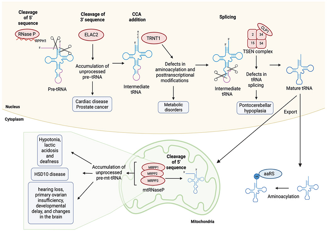
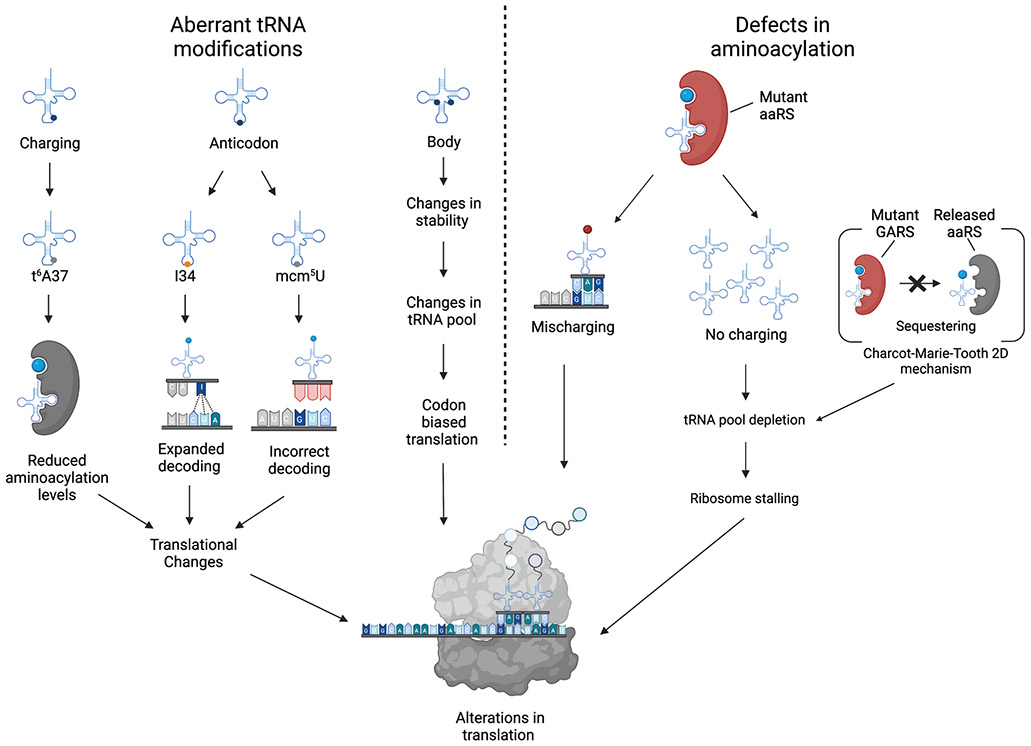
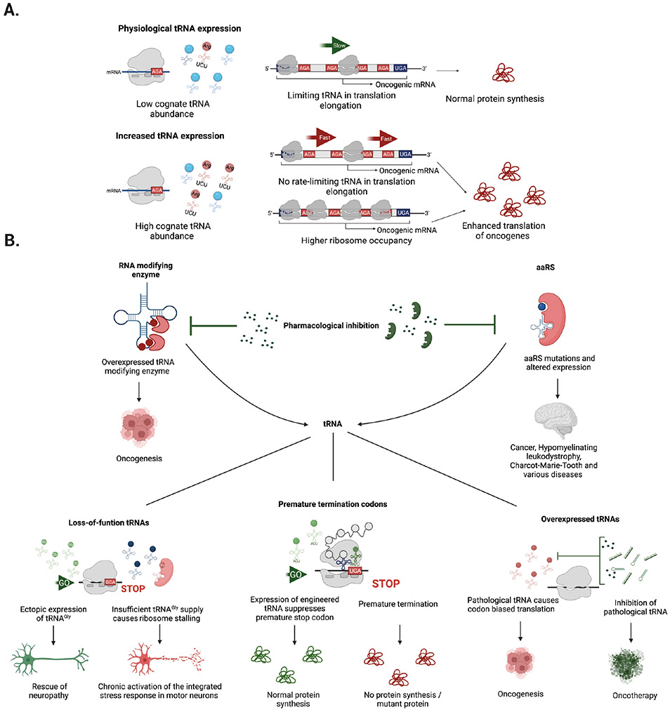
tRNAs are key adaptor molecules that decipher the genetic code during translation of mRNAs in protein synthesis. In contrast to the traditional view of tRNAs as ubiquitously expressed housekeeping molecules, awareness is now growing that tRNA-encoding genes display tissue-specific and cell type-specific patterns of expression, and that tRNA gene expression and function are both dynamically regulated by post-transcriptional RNA modifications. Moreover, dysregulation of tRNAs, mediated by alterations in either their abundance or function, can have deleterious consequences that contribute to several distinct human diseases, including neurological disorders and cancer. Accumulating evidence shows that reprogramming of mRNA translation through altered tRNA activity can drive pathological processes in a codon-dependent manner. This Review considers the emerging evidence in support of the precise control of functional tRNA levels as an important regulatory mechanism that coordinates mRNA translation and protein expression in physiological cell homeostasis, and highlights key examples of human diseases that are linked directly to tRNA dysregulation.


Figures

Figure 1.
Figure 1.. tRNA molecular structure and variance in isodecoder prevalence in eukaryotic species.
a ∣ Cloverleaf structure of a tRNA molecule. The colours denote different structural domains. b ∣ The number of tRNA isodecoders increases along the phylogenetic spectrum. Here, numbers of tRNA isodecoders are depicted as the fraction of all tRNA genes in different eukaryotes. Data for Part b were obtained from ref.
Figure 2.
Figure 2.. RNA polymerase III involvement in disease.
a ∣ Structure of RNA polymerase III (Pol III) (Protein Data Bank entry 7AST). The colours identify Pol III subunits that harbour pathological mutations in humans. b ∣ The regulatory effect of oncoproteins (red) and tumour suppressors (yellow) on Pol III-mediated transcription of tRNA genes. The three subunits of TFIIIB: TATA-box-binding protein (TBP), B double prime 1 (BDP1), and B-related factor-1 (BRF1), are shown interacting with TFIIIC. mTORC1 indirectly stimulates Pol III transcription via inhibition of MAF1, a negative regulator of Pol III. Ras small GTPases activate their downstream effector extracellular signal-regulated kinase (ERK), which induces BRF1 expression. Rb binds directly to BRF1, which inhibits Pol III recruitment. NOTCH1 stimulates tRNAVal transcription both directly and via targeting MYC. MYC directly interacts with BRF1 and activates the histone-modifying enzymes transformation and transcription domain-associated protein (TRRAP) and GCN5. P53 directly inhibits TBP and Pol III recruitment. Part a is adapted from ref.
Figure 3.
Figure 3.. Epigenetic mechanisms and sequence variability might influence nuclear tRNA expression.
a ∣ Epigenetic mechanisms might regulate tRNA expression. For example, methylation of tRNA genes could block DNA polymerase III (Pol III) transcription. Similarly, histone marks (such as K3K27 acetylation) and overall nucleosome occupancy could influence the accessibility of chromatin at tRNA loci. b ∣ tRNA genes show high variability in their flanking regions and these hyper-variable regions overlap with Pol III occupancy and transcription start sites (TSSs). Top plot shows the frequency of single nucleotide polymorphisms (SNPs) for active and inactive tRNA loci. Bottom plot represents the distribution of Pol III occupation relative to each position within the tRNA gene and its flanking regions. The acceptor stem (blue), D-stem (red), anticodon stem (green), and T-stem (orange) are highlighted in the 2D tRNA structure for both plots. Flanking tRNA regions containing relatively high mutation rates can overlap with Pol III TSSs, which raises the important question of whether variable flanking regions influence tRNA expression. c ∣ Mutations in nuclear encoded tRNAs are rare, but one example is a mutation in the acceptor stem region (red) of the tRNASec gene that causes reduced U34 modifications and a deficit in stress-related translation of selenoproteins. Patients with mutant tRNASec experience low plasma selenium levels, abdominal pain, fatigue, and muscle weakness. Part b is adapted from ref (top graph) and ref (bottom graph).
Figure 4.
Figure 4.. tRNA maturation and splicing defects in disease.
tRNA maturation involves the removal of 5' leader and 3' trailer sequences, CCA addition, and in some instances splicing of a short intron. Subsequently the mature tRNA is exported to the cytoplasm for aminoacylation (AA) by aminoacyl tRNA synthetases (aaRS). Some nuclear encoded tRNAs are imported into mitochondria. Mutations in proteins involved in the maturation of tRNAs (red) can lead to disease (shaded boxes). Levels of ribonuclease P (RNAse P) and ribonuclease P RNA component 1 (RPPH1) enzymes are increased in cancer but the physiological relevance of these alterations remains unknown.
Figure 5.
Figure 5.. Defects in tRNA modifications or aminoacylation influence translation.
Translation elongation factors (principally eIF1α) capture aminoacylated tRNAs (with the exceptions of initiator tRNAMet, which requires eIF218, and tRNASec, which requires eIFSec19) and deliver them to ribosomes to be utilized in the synthesis of proteins. Inside the A-site of the ribosome, the tRNA recognizes cognate codon sequences by forming hydrogen bonds with its anticodon triplet at positions 34, 35, and 36. Codon–anticodon interactions in positions 35 and 36 follow Watson–Crick base pairing, whereas those at position 34 sometimes do not (wobble pairing). Thus, the canonical function of tRNAs as adapter molecules during protein synthesis is influenced by changes in tRNA modifications. Absence of the mcm5U modification in the anticodon results in incorrect decoding, whereas I34 modifications in the anticodon expand the tRNA decoding capabilities. Furthermore, tRNAs lacking t6A37 modifications leads to reduced aminoacylation levels. Moreover, defects in aminoacyl tRNA synthetases (aaRS) can cause mischarging (red circle), resulting in either incorporation of an incorrect amino acid or the absence of amino acid charging followed by tRNA degradation, depletion of the corresponding tRNA, and ribosome stalling at the cognate codon. The best-understood example is Charcot–Marie–Tooth disease type 2D, in which mutant glycyl-tRNA synthetase (GARS) fails to release bound tRNAGly, thereby sequestering it and leading to depletion of functional tRNAGly .
Figure 6.
Figure 6.. Alterations in the tRNA pool can drive disease and offer avenues for therapeutic intervention.
a ∣ Dysregulation of the levels of specific tRNAs leads to codon-biased translation. During steady state, the abundance of certain tRNAs could be a rate-limiting factor for the translation of oncogenic mRNAs enriched in their cognate codon (AGA highlighted in red), which restricts the translation rate and results in normal protein synthesis. Upregulation of the corresponding tRNA (red modified tRNAArg(TCT) versus other tRNAs (blue) removes this restriction, raising translation rates and increasing the production of oncogenic protein. This mechanism can result from increased ribosome occupancy or faster (enhanced) translation of mRNAs containing its cognate codon (red AGA). b ∣ Potential avenues for therapeutic intervention include pharmacological inhibition of tRNA modifying enzymes or aminoacyl tRNA synthetases (aaRSs) using small molecules or biological agents. These approaches could correct dysfunctional tRNA levels in cancer and various neurological diseases. Similarly, modulation of the expression of downstream tRNA effectors, either by ectopic restoration (green tRNA molecules) or inhibition of overexpressed tRNAs (red) might offer new strategies to treat diseases caused by altered levels of functional tRNAs. Potential approaches include pharmacological inhibition of pathological tRNAs by small molecules, RNA interference (RNAi), or antisense oligonucleotides (green). Another approach is the expression of anticodon engineered tRNAs to suppress premature termination codons and achieve normal protein synthesis.

References

    1. Hoagland MB, Stephenson ML, Scott JF, Hecht LI & Zamecick PC A soluble ribonucleic acid intermediate in protein synthesis. J. Biol. Chem 231, 241–257 (1958).

      This paper described the discovery of tRNAs.

    1. Holley RW et al. Structure of a ribonucleic acid. Science 147, 1462–1465 (1965).

      This paper described the structure of tRNAs and the finding of a modified base (inosine).

    1. Crick FHC Codon-anticodon pairing: the wobble hypothesis. J. Mol. Biol 19, 548–555 (1966).

      This paper described the possibility of non-canonical (wobble) pairing of the anticodon loop with its cognate codon sequence on mRNAs.

    1. Chou H-J, Donnard E, Gustafsson HT, Garber M & Rando OJ Transcriptome-wide analysis of roles for tRNA modifications in translational regulation. Mol. Cell 68, 978–992.e4 (2017). - PMC - PubMed
    1. De Crécy-Lagard V. et al. Matching tRNA modifications in humans to their known and predicted enzymes. Nucleic Acids Res. 47, 2143–2159 (2019). - PMC - PubMed

Publication types