

Review
doi: 10.1177/03000605221105003.
Prolonged durability of extensive contiguous spinal metastasis stabilization in non-small cell lung cancer patients receiving targeted therapy: two case reports and a literature review
Affiliations
- PMID: 35681249
- PMCID: PMC9189544
- DOI: 10.1177/03000605221105003
Item in Clipboard
Review
Prolonged durability of extensive contiguous spinal metastasis stabilization in non-small cell lung cancer patients receiving targeted therapy: two case reports and a literature review
J Int Med Res.
2022 Jun.
Abstract
In this report, we present two cases of contiguous spinal metastatic disease in non-small cell lung cancer patients who achieved prolonged survival and stable spinal fixation after treatment with EGFR TKIs.
Keywords: Spinal metastasis; epidermal growth factor tyrosine kinase inhibitor; non-small cell lung cancer; quality of life; spinal cord compression; surgery; targeted therapy.
Conflict of interest statement
Figures

Case 1: Chest computed tomography (CT) (upper images: coronal views; lower images: axial views) (a) Initial CT scan showing a 1.7-cm spiculated mass in the left upper lung suggesting primary lung cancer, with multiple lung and lymph node metastases. (b) Four months after treatment with gefitinib, decreases in the sizes of the primary lung mass, metastatic pulmonary nodules, and lymph nodes are noted and (c) At 26 months, the patient developed EGFR TKI resistance. Disease progression is evident by the increased sizes of the primary lung mass and metastatic pulmonary nodules. EGFR TKI, epidermal growth factor receptor tyrosine kinase inhibitor.
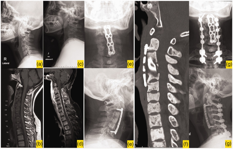
Case 1: Imaging of spinal metastatic disease (a) Initial plain radiograph (lateral view) showing an osteolytic lesion with minimal vertebral height loss in the C4 vertebra. (b) T1-weighted MRI (sagittal view) showing contiguous spinal metastasis from C2 to T6. (c) Plain radiograph (lateral view) 1 month after initial presentation showing that bony destruction has progressed, with increasing collapse of the C4 vertebra. (d) T2-weighted MRI (sagittal view) showing spinal cord compression at the C4 level and spinal metastasis from C2–T6. (e) Postoperative plain radiographs (upper image, anteroposterior view; lower image: lateral view) after anterior cervical corpectomy of C3–C5 with a fibular strut autograft and C2–C6 anterior plating. (f) Postoperative CT image (sagittal view) after systemic treatment with EGFR TKIs, EBRT, and zoledronic acid. An osteoblastic reaction is visible along the previously lytic spine and (g) Plain radiographs (upper image: anteroposterior view; lower image: lateral view) at 39 months, after posterior supplemental fixation at 7 months. The spinal construct is still durable. MRI, magnetic resonance image; EGFR TKI, epidermal growth factor receptor tyrosine kinase inhibitor; EBRT, external beam radiation therapy.

Case 2: Chest computed tomography (CT) (upper images: coronal views; lower images: axial views) (a) Initial CT images showing a large mass in the left upper lung, with paratracheal lymph node metastasis. (b) Five months after treatment with gefitinib, a reduction in the size of the primary lung mass size is observed and (c) At 24 months, the patient developed EGFR TKI resistance, which resulted in increases in the sizes of the index lung mass and paratracheal lymph nodes, and new lung metastases. EGFR TKI, epidermal growth factor receptor tyrosine kinase inhibitor.

Case 2: Imaging of spinal metastatic disease (a) Initial plain radiographs (upper image: anteroposterior view; lower image: lateral view) showing subtle osteolytic lesions along the subaxial cervical vertebrae. (b) T1- (upper image) and T2- (lower image) weighted sagittal MRI showing contiguous spinal metastasis from C4 to T6, with spinal cord compression at T2. (c) Postoperative plain radiographs (upper image: anteroposterior view; lower image: lateral view) after decompressive laminectomy at T1–T2 with transpedicular decompression at T2 and T1–T5 spinal instrumentation. A transverse process hook was applied at T1 owing to bony destruction. (d) Sagittal CT image, 5 months after treatment showing that an osteoblastic bone reaction is visible in the previously lytic regions and (e) Plain radiographs (upper image: anteroposterior view; lower image: lateral view) at 26 months showing that the spinal fixation construct is stable, without signs of implant loosening. MRI, magnetic resonance image.
References
-
- Aydinli U, Ozturk C, Bayram S, et al.. Evaluation of lung cancer metastases to the spine. Acta Orthop Belg 2006; 72: 592–597. - PubMed
-
- Fehlings MG, Nater A, Tetreault L, et al.. Survival and clinical outcomes in surgically treated patients with metastatic epidural spinal cord compression: results of the prospective multicenter AOSpine study. J Clin Oncol 2016; 34: 268–276. - PubMed
-
- Kobayashi K, Ando K, Nakashima H, et al.. Prognostic factors in the new Katagiri scoring system after palliative surgery for spinal metastasis. Spine (Phila Pa 1976) 2020; 45: E813–E819. - PubMed
Publication types
MeSH terms
Substances
LinkOut - more resources
Full Text Sources
Medical
Research Materials
Miscellaneous
