Review
. 2022 May 27;11(11):1757.
doi: 10.3390/cells11111757.

Reovirus Activated Cell Death Pathways

Affiliations
Review

Reovirus Activated Cell Death Pathways

Carly DeAntoneo et al. Cells. .

Abstract

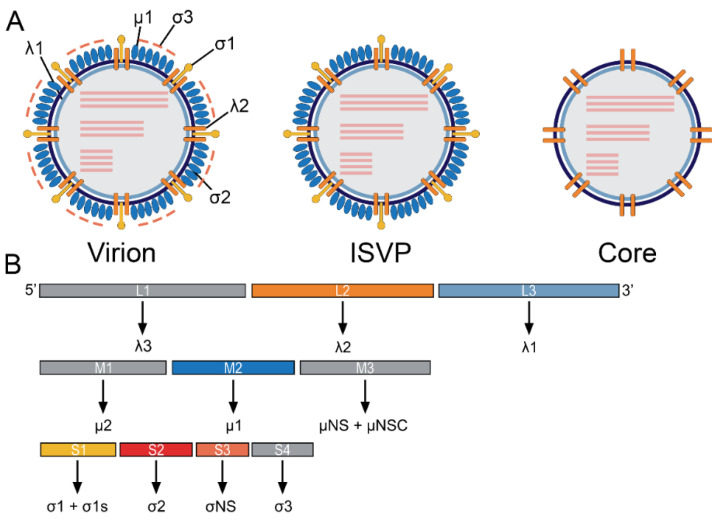
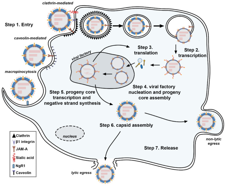
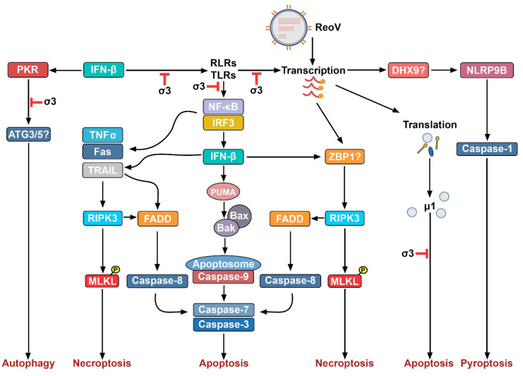
Mammalian orthoreoviruses (ReoV) are non-enveloped viruses with segmented double-stranded RNA genomes. In humans, ReoV are generally considered non-pathogenic, although members of this family have been proven to cause mild gastroenteritis in young children and may contribute to the development of inflammatory conditions, including Celiac disease. Because of its low pathogenic potential and its ability to efficiently infect and kill transformed cells, the ReoV strain Type 3 Dearing (T3D) is clinical trials as an oncolytic agent. ReoV manifests its oncolytic effects in large part by infecting tumor cells and activating programmed cell death pathways (PCDs). It was previously believed that apoptosis was the dominant PCD pathway triggered by ReoV infection. However, new studies suggest that ReoV also activates other PCD pathways, such as autophagy, pyroptosis, and necroptosis. Necroptosis is a caspase-independent form of PCD reliant on receptor-interacting serine/threonine-protein kinase 3 (RIPK3) and its substrate, the pseudokinase mixed-lineage kinase domain-like protein (MLKL). As necroptosis is highly inflammatory, ReoV-induced necroptosis may contribute to the oncolytic potential of this virus, not only by promoting necrotic lysis of the infected cell, but also by inflaming the surrounding tumor microenvironment and provoking beneficial anti-tumor immune responses. In this review, we summarize our current understanding of the ReoV replication cycle, the known and potential mechanisms by which ReoV induces PCD, and discuss the consequences of non-apoptotic cell death-particularly necroptosis-to ReoV pathogenesis and oncolysis.

Keywords: MLKL; RIPK3; ZBP1; apoptosis; necroptosis; oncolysis; reovirus.


Conflict of interest statement

The authors declare no conflict of interest.

Figures

Figure 1
Figure 1
ReoV structure and genome organization. (A) Schematic depiction of the mature virion, containing all virally encoded proteins (left); the ISVP, a replication intermediate which has shed the σ3 protein (middle); and the viral core, lacking σ3, σ1, and µ1 (right). Viral proteins are indicated by black lines. Viral dsRNA genome segments are portrayed as pink lines. (B) ReoV genome comprises 10 dsRNA segments, depicted as rectangles. The proteins encoded by each segment are portrayed below.
Figure 2
Figure 2
Reovirus Life Cycle. ReoV enters cells (Step 1) following binding of σ1 protein to host cell receptor, such as JAM-A and, in some cases, sialic acids. Clathrin-mediated endocytosis is the most common entry mechanism, although caveolin-mediated endocytosis and macropinocytosis is also possible. The virions are then shuttled within endosomes. Proteolysis degrades the outer σ3 protein and cleavage of the unveiled µ1C into fragments δ and φ, leading to formation of the ISVP. The µ1C cleavage fragments, along with µ1N, form pores within the endosomal membrane, depositing the viral core now lacking both µ 1 and σ1. Transcription by the RdRp λ3 occurs within the viral core underneath channels formed by λ2 (Step 2). Capping of mRNA is mediated by λ2. Viral mRNA is exported from the viral core and into the cytoplasm, where translation occurs (Step 3). Viral proteins mediate nucleation of viral factories, where progeny cores begin to self-assemble (Step 4). Negative strand synthesis occurs within progeny cores, forming nascent viral genomes (Step 5). Progeny core transcription occurs, and outer capsid proteins begin to assemble around progeny cores (Step 6). Finally, progeny virons leave the cell, either via lytic or non-lytic egress (Step 7).
Figure 3
Figure 3
Programmed Cell Death Pathways activated by ReoV. This figure illustrates four potential programmed cell death pathways activated by ReoV infection. Potential dsRNA sensors in these pathways are indicated by question marks.

References

    1. Dermody T.S., Parker J.S., Barbara S. Orthoreoviruses. In: Knipe D.M., Howley P.M., editors. Fields Virology. 6th ed. Volume 2. Lippincott Williams & Wilkins; Philadelphia, PA, USA: 2013. pp. 1304–1346.
    1. Bouziat R., Hinterleitner R., Brown J.J., Stencel-Baerenwald J.E., Ikizler M., Mayassi T., Meisel M., Kim S.M., Discepolo V., Pruijssers A.J., et al. Reovirus infection triggers inflammatory responses to dietary antigens and development of celiac disease. Science. 2017;356:44–50. doi: 10.1126/science.aah5298. - DOI - PMC - PubMed
    1. Sutherland D.M., Aravamudhan P., Dietrich M.H., Stehle T., Dermody T.S. Reovirus Neurotropism and Virulence Are Dictated by Sequences in the Head Domain of the Viral Attachment Protein. J. Virol. 2018;92:e00974-18. doi: 10.1128/JVI.00974-18. - DOI - PMC - PubMed
    1. Gauvin L., Bennett S., Liu H., Hakimi M., Schlossmacher M., Majithia J., Brown E.G. Respiratory infection of mice with mammalian reoviruses causes systemic infection with age and strain dependent pneumonia and encephalitis. Virol. J. 2013;10:67. doi: 10.1186/1743-422X-10-67. - DOI - PMC - PubMed
    1. Konopka-Anstadt J.L., Mainou B.A., Sutherland D.M., Sekine Y., Strittmatter S.M., Dermody T.S. The Nogo receptor NgR1 mediates infection by mammalian reovirus. Cell Host Microbe. 2014;15:681–691. doi: 10.1016/j.chom.2014.05.010. - DOI - PMC - PubMed

Publication types

Substances