. 2022 May 27;14(11):2665.
doi: 10.3390/cancers14112665.

Ficus dubia Latex Extract Induces Cell Cycle Arrest and Apoptosis by Regulating the NF-κB Pathway in Inflammatory Human Colorectal Cancer Cell Lines

Affiliations

Ficus dubia Latex Extract Induces Cell Cycle Arrest and Apoptosis by Regulating the NF-κB Pathway in Inflammatory Human Colorectal Cancer Cell Lines

Rentong Hu et al. Cancers (Basel). .

Abstract

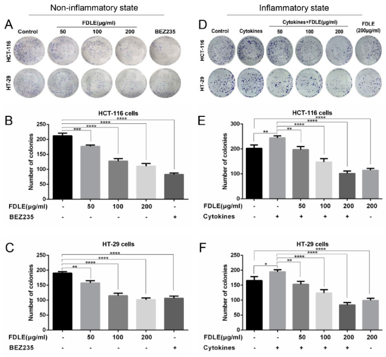
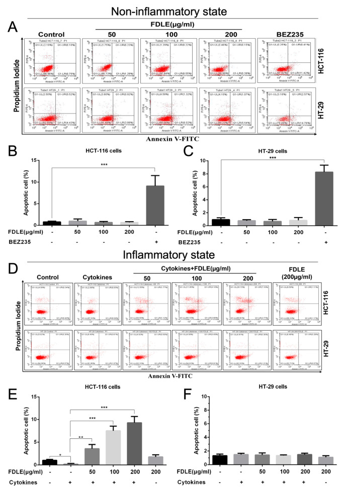
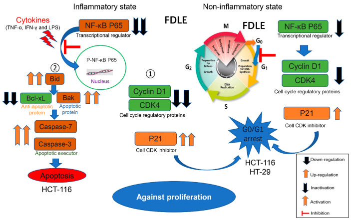
Colorectal cancer is one of the most diagnosed cancers that is associated with inflammation. Ficus dubia latex is recognized as a remedy with various therapeutic effects in traditional medicine, including anti-inflammatory and antioxidant activity. The present study aims to compare the anti-tumor activity of Ficus dubia latex extract (FDLE) against HCT-116 and HT-29 human colorectal cancer cell lines in normal and inflammatory condition and explore its mechanism of action. FDLE exhibited remarkable antiproliferative activity against HCT-116 and HT-29 colorectal cancer cell lines in both conditions using MTT and colony formation assays and more effective anti-proliferation was observed in inflammatory condition. Mechanistically, FDLE induced cell cycle arrest at G0/G1 phase by down-regulating NF-κB, cyclin D1, CDK4 and up-regulatingp21 in both cell in normal condition. In inflammatory condition, FDLE not only exhibited stronger induction of cell cycle arrest in both cells by down-regulating NF-κB, cyclin D1, CDK4 and down-regulating p21, but also selectively induced apoptosis in HCT-116 cells by down-regulating NF-κB and Bcl-xl and up-regulating Bid, Bak, cleaved caspase-7 and caspase-3 through stronger ability to regulate these proteins. Our results demonstrated that the phytochemical agent in the latex of Ficus dubia could potential be used for treatment and prevention of human colorectal cancer, especially in inflammation-induced hyperproliferation progression.

Keywords: Ficus dubia latex extract (FDLE); NF-κB pathway; apoptosis; cell cycle arrest; colorectal cancer; inflammation; proliferation.


Conflict of interest statement

The authors declare no conflict of interest.

Figures

Figure 1
Figure 1
The experimental design of non-inflammatory and inflammatory models.
Figure 2
Figure 2
The effects of FDLE on cell viability of colorectal cancer cell lines (HCT-116 and HT-29) and normal mouse embryonic fibroblast cell (NIH3T3) in non-inflammatory and inflammatory states by MTT. The cells were treated with various concentrations of FDLE for 24, 48 and 72 h. The cell viability was calculated comparing to untreated control cells after 24, 48 and 72 h of incubation. (AC) The cell viability of HCT-116, HT-29 and NIH3T3 cells in non-inflammatory state. (DF) The cell viability of HCT-116, HT-29 and NIH3T3 cells in inflammatory state. The results were represented as mean ± SD of three independent experiments, * p < 0.05, ** p < 0.01, *** p < 0.001, **** p < 0.0001.
Figure 3
Figure 3
The effects of FDLE on growth inhibition in non-inflammatory and inflammatory HCT-116 and HT-29 colorectal cancer cells by colony formation assay. (AC) Colony number of non-inflammatory HCT-116 and HT-29 cells treated with three doses (50, 100 and 200 µg/mL) of FDLE for 48 h. (DF) Colony number of inflammatory HCT-116 and HT-29 cells treated with three doses (50, 100 and 200 µg/mL) of FDLE for 48 h after adding a mixture of cytokines (TNF-α, IFN-γ and LPS each 10 ng/mL) for 2 h. The cells were washed by PBS and cultured for 7 days. The results were represented as mean ± SD of three independent experiments, * p < 0.05, ** p < 0.01, *** p < 0.001, **** p < 0.0001.
Figure 4
Figure 4
The effects of FDLE on cell cycle arrest in non-inflammatory and inflammatory HCT-116 and HT-29 colorectal cancer cells by flow cytometry. (AC) Representative cell cycle distribution of non-inflammatory HCT-116 and HT-29 cells treated with three doses (50, 100 and 200 µg/mL) of FDLE for 48 h. (DF) Representative cell cycle distribution of inflammatory HCT-116 and HT-29 cells treated with three doses (50, 100 and 200 µg/mL) of FDLE for 48 h after adding a mixture of cytokines (TNF-α, IFN-γ and LPS each 10 ng/mL) for 2 h. The results were represented as mean ± SD of three independent experiments, * p < 0.05, ** p < 0.01, *** p < 0.001, **** p < 0.0001.
Figure 5
Figure 5
The effects of FDLE on NF-κB-mediated cell cycle proteins in non-inflammatory and inflammatory HCT-116 and HT-29 colorectal cancer cell line. (A,B) Western blot analysis of NF-κB-mediated proteins related to cell cycle in non-inflammatory HCT-116 and HT-29 cells treated with three doses (50, 100 and 200 µg/mL) of FDLE for 48 h. (C,D) Western blot analysis of NF-κB-mediated proteins related to cell cycle in inflammatory HCT-116 and HT-29 cells treated with three doses (50, 100 and 200 µg/mL) of FDLE for 48 h after adding a mixture of cytokines (TNF-α, IFN-γ and LPS each 10 ng/mL) for 2 h. Densitometric quantification was performed by image software. The results were represented as mean ± SD of three independent experiments, * p < 0.05, ** p < 0.01, *** p < 0.001, **** p < 0.0001. The whole western blots are shown in File S1.
Figure 6
Figure 6
The effects of FDLE on apoptosis induction in non-inflammatory and inflammatory HCT-116 and HT-29 colorectal cancer cell lines. Apoptosis includes early apoptosis (LR) and late apoptosis (UR). (AC) Apoptosis rate of non-inflammatory HCT-116 and HT-29 cells treated with three doses (50, 100 and 200 µg/mL) of FDLE for 48 h. (DF) Apoptosis rate of inflammatory HCT-116 and HT-29 cells treated with three doses (50, 100 and 200 µg/mL) of FDLE for 48 h after adding a mixture of cytokines (TNF-α, IFN-γ and LPS each 10 ng/mL) for 2 h. The results were represented as mean ± SD of three independent experiments, * p < 0.05, ** p < 0.01, *** p < 0.0001.
Figure 7
Figure 7
The effects of FDLE on NF-κB-mediated expression of apoptotic proteins and activation of caspase enzymes in non-inflammatory and inflammatory HCT-116 and HT-29 colorectal cancer cell lines by Western blot. (A,B) The expression of apoptotic proteins and the activation of caspase enzymes in non-inflammation HCT-116 and HT-29 cells untreated and treated with three doses (50, 100 and 200 µg/mL) of FDLE or BEZ235 for 48 h. (C,D) The expression of apoptotic proteins and the activation of caspase enzymes in inflammation HCT-116 and HT-29 cells untreated and treated with three doses (50, 100 and 200 µg/mL) of FDLE for 48 h after adding cytokines (TNF-α, IFN-γ and LPS each 10 ng/mL) for 2 h. Densitometric quantification was performed by image software. The results were represented as mean ± SD of three independent experiments, * p < 0.05, ** p < 0.01, *** p < 0.001, **** p < 0.0001. The whole western blots are shown in File S1.
Figure 8
Figure 8
The different anti-proliferative mechanisms of FDLE on HCT-116 and HT-29 human colorectal cancer cell lines in non-inflammatory and inflammatory conditions.

References

    1. Long A.G., Lundsmith E.T., Hamilton K.E. Inflammation and colorectal cancer. Curr. Colorectal Cancer Rep. 2017;13:341–351. doi: 10.1007/s11888-017-0373-6. - DOI - PMC - PubMed
    1. Park C.H., Eun C.S., Han D.S. Intestinal microbiota, chronic inflammation, and colorectal cancer. Intest. Res. 2018;16:338. doi: 10.5217/ir.2018.16.3.338. - DOI - PMC - PubMed
    1. Ieda T., Tazawa H., Okabayashi H., Yano S., Shigeyasu K., Kuroda S., Ohara T., Noma K., Kishimoto H., Nishizaki M. Visualization of epithelial-mesenchymal transition in an inflammatory microenvironment–colorectal cancer network. Sci. Rep. 2019;9:16378. doi: 10.1038/s41598-019-52816-z. - DOI - PMC - PubMed
    1. Yoon S.-W. Systemic inflammatory response as a prognostic factor in patients with cancer. J. Korean Tradit. Oncol. 2012;17:1–7.
    1. Ullman T.A., Itzkowitz S.H. Intestinal inflammation and cancer. Gastroenterology. 2011;140:1807–1816.e1. doi: 10.1053/j.gastro.2011.01.057. - DOI - PubMed