. 2022 Jun 1;14(11):2749.
doi: 10.3390/cancers14112749.

Targeting Immunosuppressive Tumor-Associated Macrophages Using Innate T Cells for Enhanced Antitumor Reactivity

Affiliations

Targeting Immunosuppressive Tumor-Associated Macrophages Using Innate T Cells for Enhanced Antitumor Reactivity

Yan-Ruide Li et al. Cancers (Basel). .

Abstract

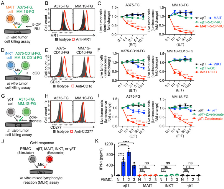
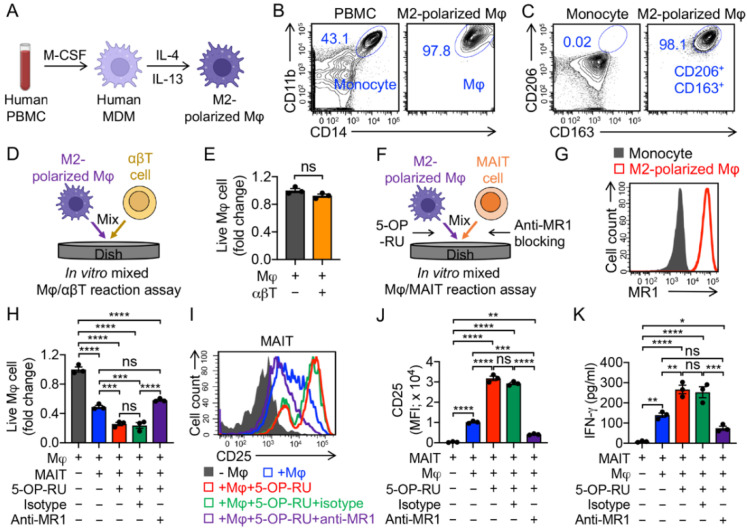
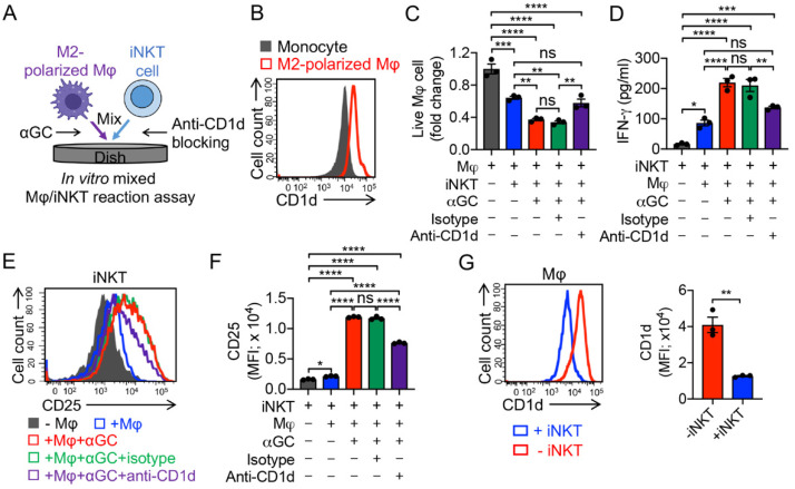
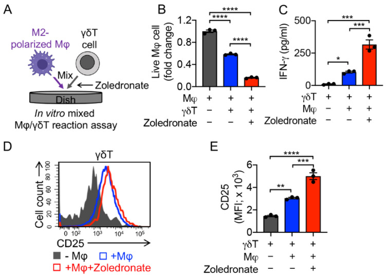
The field of T cell-based and chimeric antigen receptor (CAR)-engineered T (CAR-T) cell-based antitumor immunotherapy has seen substantial developments in the past decade; however, considerable issues, such as graft-versus-host disease (GvHD) and tumor-associated immunosuppression, have proven to be substantial roadblocks to widespread adoption and implementation. Recent developments in innate immune cell-based CAR therapy have opened several doors for the expansion of this therapy, especially as it relates to allogeneic cell sources and solid tumor infiltration. This study establishes in vitro killing assays to examine the TAM-targeting efficacy of MAIT, iNKT, and γδT cells. This study also assesses the antitumor ability of CAR-engineered innate T cells, evaluating their potential adoption for clinical therapies. The in vitro trials presented in this study demonstrate the considerable TAM-killing abilities of all three innate T cell types, and confirm the enhanced antitumor abilities of CAR-engineered innate T cells. The tumor- and TAM-targeting capacity of these innate T cells suggest their potential for antitumor therapy that supplements cytotoxicity with remediation of tumor microenvironment (TME)-immunosuppression.

Keywords: cancer immunotherapy; gamma delta T (γδT) cells; innate T cell; invariant natural killer T (iNKT) cell; mucosal-associated invariant T (MAIT) cell; tumor microenvironment (TME); tumor-associated macrophage.


Conflict of interest statement

L.Y. is a scientific advisor to AlzChem and Amberstone Biosciences, and a co-founder, stockholder, and advisory board member of Appia Bio. None of the declared companies contributed to or directed any of the research reported in this article. The present authors declare no competing interests.

Figures

Figure 1
Figure 1
Generation of human peripheral blood mononuclear cell (PBMC)-derived T cells. (A,C,E,G) Diagram outlining the generation of PBMC-derived conventional αβ T (αβT), mucosal-associated invariant T (MAIT), invariant natural killer T (iNKT), and gamma delta T (γδT) cells. (B,D,F,H) Fluorescence-activated cell sorting (FACS) detection of the generation and CD4/CD8 co-receptor expression of αβT, MAIT, iNKT, and γδT cells. Representative of over five experiments.
Figure 2
Figure 2
In vitro antitumor tumor efficacy and safety study of PBMC-derived T cells. (AC) In vitro killing of human A375 melanoma and MM.1S multiple myeloma cells by MAIT cells. Both tumor cell lines were engineered to express firefly luciferase and green florescence protein (FG) dual reporters. Moreover, 5-(2-oxopropylideneamino)-6-d-ribitylaminouracil (5-OP-RU) was added to activate MAIT cells. (A) Experimental design. (B) FACS detection of MR1 on A375-FG and MM.1S-FG cells. (C) Tumor killing data at 24 h (n = 4). (DF) In vitro killing of A375 and MM.1S cells by iNKT cells. Both tumor cell lines were engineered to express human CD1d as well as FG dual reporters, and α-galactosylceramide (αGC) was added to activate iNKT cells. (D) Experimental design. FACS detection of CD1d on A375-CD1d-FG and MM.1S-CD1d-FG cells. (F) Tumor killing data at 24 h (n = 4). (GI) In vitro killing of A375 and MM.1S cells by γδT cells. Zoledronate was added to activate γδT cells. (G) Experimental design. (H) FACS detection of CD277 on A375-FG and MM.1S- FG cells. (I) Tumor killing data at 24 h (n = 4). (J,K) Studying the graft-versus-host (GvH) response of PBMC-derived T cells using an in vitro mixed lymphocyte reaction (MLR) assay. (J) Experimental design. PBMCs from three different healthy donors were used as stimulator cells. (K) ELISA analyses of IFN-γ secretion at day 4 (n = 3). N, no stimulator cells. Representative of three experiments. Data are presented as the mean ± SEM. ns, not significant, ** p < 0.01, *** p < 0.001, **** p < 0.0001, by one-way ANOVA.
Figure 3
Figure 3
In vitro targeting of immunosuppressive macrophages by PBMC-derived MAIT cells. (AC) In vitro generation and polarization of human monocyte-derived M2 macrophages. (A) Experimental design. M-CSF, macrophage colony-stimulating factor; MDM, monocyte-derived macrophage; Mφ, macrophage. (B) FACS detection of CD11b and CD14 on M2-polarized macrophages. Healthy donor PBMCs were included as a staining control. (C) FACS detection of M2 macrophage markers (i.e., CD163 and CD206) on M2-polarized macrophages. Monocytes were included as a control. (D,E) Studying macrophage targeting by αβ T cells using an in vitro mixed macrophage/αβ T cell (Mφ/αβT) reaction assay. (D) Experimental design. (E) FACS analysis of live macrophages 24 h after co-culturing with αβ T cells. Live cells were identified as e506CD14+CD11b+ (n = 3). (FK) Studying macrophage targeting by MAIT cells using an in vitro mixed macrophage/MAIT cell (Mφ/MAIT) reaction assay; 5-OP-RU was added to activate MAIT cells. (F) Experimental design. (G) FACS detection of MR1 on monocytes and M2-polarized macrophages. (H) FACS analysis of live macrophages 24 h after co-culturing with MAIT cells. (I) FACS detection of CD25 expression on MAIT cells. (J) Quantification of I (n = 3). (K) ELISA analysis of IFN-γ secretion by MAIT cells in the supernatants of various mixed cell cultures (n = 3). Representative of three experiments. Data are presented as the mean ± SEM. ns, not significant, * p < 0.05, ** p < 0.01, *** p < 0.001, **** p < 0.0001, by Student’s t test (E), or by one-way ANOVA (H,J,K).
Figure 4
Figure 4
In vitro targeting of immunosuppressive macrophages by PBMC-derived iNKT cells. (A) Experimental design. αGC was added to activate iNKT cells. (B) FACS detection of CD1d on monocytes and M2-polarized macrophages. (C) FACS analysis of live macrophages 24 h after co-culturing with iNKT cells. (D) ELISA analysis of IFN-γ secretion by iNKT cells in the supernatants of various mixed cell cultures (n = 3). (E) FACS detection of CD25 expression on iNKT cells. (F) Quantification of E (n = 3). (G) FACS analysis of CD1d expression on macrophages with or without co-culturing with iNKT cells. Representative of three experiments. Data are presented as the mean ± SEM. ns, not significant, * p < 0.05, ** p < 0.01, *** p < 0.001, **** p < 0.0001, by Student’s t test (G), or by one-way ANOVA (C,D,F).
Figure 5
Figure 5
In vitro targeting of immunosuppressive macrophages by PBMC-derived γδT cells. (A) Experimental design. Zoledronate was added to activate γδT cells. (B) FACS analysis of live macrophages 24 h after co-culturing with γδT cells. (C) ELISA analysis of IFN-γ secretion by γδT cells in the supernatants of various mixed cell cultures (n = 3). (D) FACS detection of CD25 expression on γδT cells. (E) Quantification of D (n = 3). Representative of three experiments. Data are presented as the mean ± SEM. ns, not significant, * p < 0.05, ** p < 0.01, *** p < 0.001, **** p < 0.0001, by one-way ANOVA.
Figure 6
Figure 6
Targeting TAMs by mesothelin-targeting CAR-engineered MAIT (MCAR-MAIT) cells using an ex vivo 3D TME mimicry culture. (A,B) Diagram outlining the generation of MCAR-engineered αβT (MCAR-αβT) and MAIT cells. (C) FACS detection of MCAR expression on MCAR-αβT and MCAR-MAIT cells. (DF) In vitro killing of human ovarian cancer OVCAR3 cells by MCAR-MAIT cells. OVCAR3-FG, human OVCAR3 cell line engineered to overexpress FG. MCAR-αβT cells were included as an effector cell control. MCAR-MAIT and MCAR-αβT cells were FACS-sorted for MCAR+ cell populations. (D) Experimental design. (E) FACS detection of mesothelin (MSLN) expression on OVCAR3-FG cells. (F) Tumor killing data at 24 h (n = 4). (GM) Studying TAM targeting by MCAR-MAIT cells using an ex vivo 3D TME mimicry culture. MCAR-αβT cells were included as an effector cell control. (G) Experimental design. (H) FACS analysis of live OVCAR3-FG cells 48 h after co-culturing with MCAR-αβT cells. (n = 3). (I) FACS detection of CD25 expression on MCAR-αβT cells (n = 3). (J) FACS analysis of live macrophages 48 h after co-culturing with MCAR-αβT cells (n = 3). Live macrophages were identified as e506CD14+CD11b+. (K) FACS analysis of live OVCAR3-FG cells 48 h after co-culturing with MCAR-MAIT cells. (n = 3). (L) FACS detection of CD25 expression on MCAR-MAIT cells (n = 3). (M) FACS analysis of live macrophages 48 h after co-culturing with MCAR-MAIT cells (n = 3). Representative of three experiments. Data are presented as the mean ± SEM. ns, not significant, * p < 0.05, ** p < 0.01, *** p < 0.001, **** p < 0.0001, by Student’s t test (F,I,J), or by one-way ANOVA (H,KM).

References

    1. Labanieh L., Majzner R.G., Mackall C.L. Programming CAR-T cells to kill cancer. Nat. Biomed. Eng. 2018;2:377–391. doi: 10.1038/s41551-018-0235-9. - DOI - PubMed
    1. Kasakovski D., Xu L., Li Y. T cell senescence and CAR-T cell exhaustion in hematological malignancies. J. Hematol. Oncol. 2018;11:91. doi: 10.1186/s13045-018-0629-x. - DOI - PMC - PubMed
    1. Mo F., Mamonkin M., Brenner M.K., Heslop H.E. Taking T-Cell Oncotherapy Off-the-Shelf. Trends Immunol. 2021;42:261–272. doi: 10.1016/j.it.2021.01.004. - DOI - PMC - PubMed
    1. Li Y.-R., Zhou Y., Kim Y.J., Zhu Y., Ma F., Yu J., Wang Y.-C., Chen X., Li Z., Zeng S., et al. Development of allogeneic HSC-engineered iNKT cells for off-the-shelf cancer immunotherapy. Cell Rep. Med. 2021;2:100449. doi: 10.1016/j.xcrm.2021.100449. - DOI - PMC - PubMed
    1. Rafiq S., Hackett C.S., Brentjens R.J. Engineering strategies to overcome the current roadblocks in CAR T cell therapy. Nat. Rev. Clin. Oncol. 2020;17:147–167. doi: 10.1038/s41571-019-0297-y. - DOI - PMC - PubMed