. 2022 Jun 6;14(11):2812.
doi: 10.3390/cancers14112812.

Cancer-Associated Fibroblasts Confer Gemcitabine Resistance to Pancreatic Cancer Cells through PTEN-Targeting miRNAs in Exosomes

Affiliations

Cancer-Associated Fibroblasts Confer Gemcitabine Resistance to Pancreatic Cancer Cells through PTEN-Targeting miRNAs in Exosomes

Katherine E Richards et al. Cancers (Basel). .

Abstract

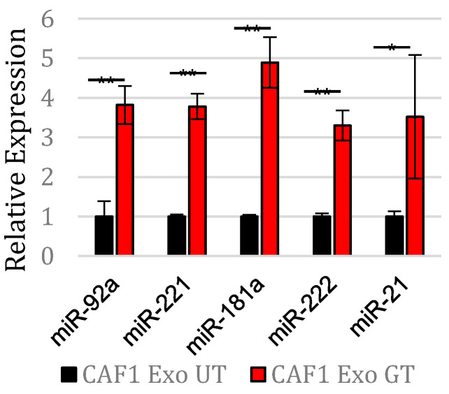
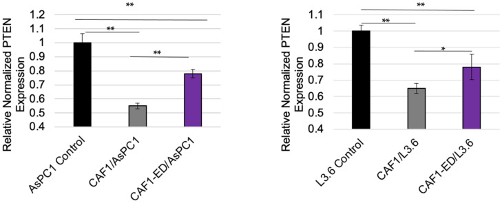
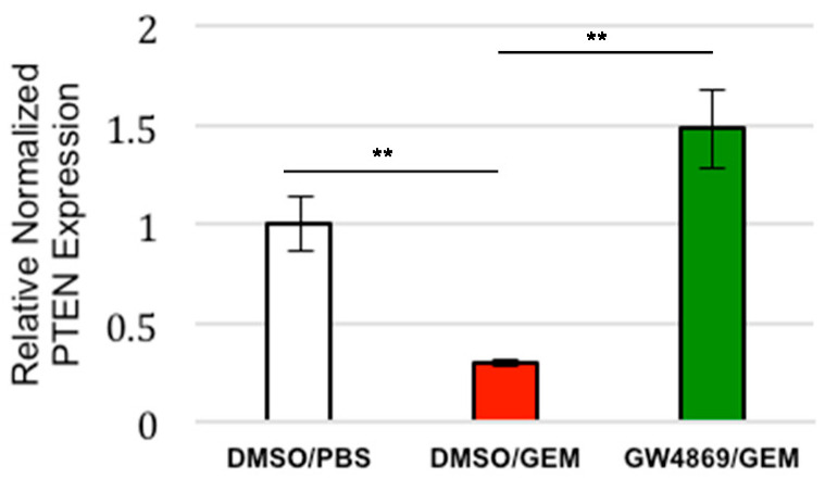
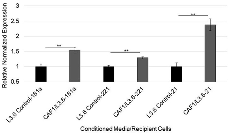
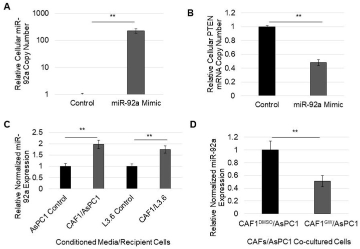
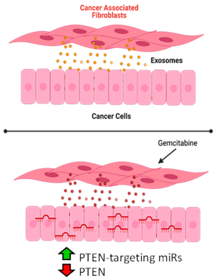
Pancreatic ductal adenocarcinoma (PDAC) is currently the third leading cause of cancer-related death in the United States. Even though the poor prognosis of PDAC is often attributed to late diagnosis, patients with an early diagnosis who undergo tumor resection and adjuvant chemotherapy still show tumor recurrence, highlighting a need to develop therapies which can overcome chemoresistance. Chemoresistance has been linked to the high expression of microRNAs (miRs), such as miR-21, within tumor cells. Tumor cells can collect miRs through the uptake of miR-containing lipid extracellular vesicles called exosomes. These exosomes are secreted in high numbers from cancer-associated fibroblasts (CAFs) within the tumor microenvironment during gemcitabine treatment and can contribute to cell proliferation and chemoresistance. Here, we show a novel mechanism in which CAF-derived exosomes may promote proliferation and chemoresistance, in part, through suppression of the tumor suppressor PTEN. We identified five microRNAs: miR-21, miR-181a, miR-221, miR-222, and miR-92a, that significantly increased in number within the CAF exosomes secreted during gemcitabine treatment which target PTEN. Furthermore, we found that CAF exosomes suppressed PTEN expression in vitro and that treatment with the exosome inhibitor GW4869 blocked PTEN suppression in vivo. Collectively, these findings highlight a mechanism through which the PTEN expression loss, often seen in PDAC, may be attained and lend support to investigations into the use of exosome inhibitors as potential therapeutics to improve the effectiveness of chemotherapy.

Keywords: PTEN; exosomes; fibroblasts; miRNAs; pancreatic cancer.


Conflict of interest statement

The authors declare no conflict of interest.

Figures

Figure 1
Figure 1
Expression of identified exosomal miRs in CAFs exposed to gemcitabine.RT-PCR normalized expression of miRs from exosomes in CAFs ex-posed to gemcitabine (Exo GT) and untreated CAFs (Exo UT). ** p < 0.01. * p < 0.05.
Figure 2
Figure 2
PTEN expression in cells where exosomes are retained or depleted from CAF-derived media. PTEN expression was analyzed via RT-PCR in AsPC1 cells (left) or L3.6 cells (right) after cultured in normal DMEM or RPMI (control), CAF-conditioned media, or exosome-depleted CAF-conditioned media (CAF1-ED) each day for four days. ** p < 0.01. * p < 0.05.
Figure 3
Figure 3
Evaluation of the effect of GEM treatment and exosome inhibition on PTEN expression in in vivo PDAC model. Mice with subcutaneous tumors (PDACs+CAFs) were treated for two weeks with PBS, Gemcitabine, or GW4869/Gemcitabine, and their PTEN mRNA levels were quantified via RT-PCR. ** p < 0.01.
Figure 4
Figure 4
Expression of PTEN-targeting miRs in cells exposed to CAF-conditioned media. L3.6 epithelial cells were cultured in DMEM or gemcitabine-treated CAF-conditioned media for four days. Expression of microRNAs within recipient L3.6 cells was thereafter quantified via RT-PCR. ** p < 0.01.
Figure 5
Figure 5
MicroRNA-92a expression in cells exposed to CAF-derived exosomes. (A) validation of mir-92a expression in a mimic-containing cell line (AsPC1). (B) PTEN mRNA levels were measured in AsPC1 cells transfected with miR-92a mimic compared to AsPC1 cells transfected with scramble negative control miR via RT-PCR. (C) miR-92a expression levels were quantified via RT-PCR within cells which received control or GEM-treated, CAF-conditioned media for four days. (D) AsPC1 cells were co-cultured underneath GEM-treated CAFs or GEM+GW4869-treated CAFs for three days. miR-92a expression within AsPC1 cells was thereafter quantified via RT-PCR. ** p < 0.01.
Figure 6
Figure 6
Schematic of proposed mechanism. CAFs routinely release exosomes which are taken up by cancer cells. CAFs treated with gemcitabine release exosomes which have increased expression of PTEN-targeting miRs. Cells that take up these exosomes have increased expression of these miRs and reduced expression of PTEN. Blockade of exosome release restores PTEN ex-pression.

Similar articles

Cited by

References

    1. Society A.C. Cancer Facts and Figures. American Cancer Society; Atlanta, GA, USA: 2022.
    1. Rahib L., Smith B.D., Aizenberg R., Rosenzweig A.B., Fleshman J.M., Matrisian L.M. Projecting cancer incidence and deaths to 2030: The unexpected burden of thyroid, liver, and pancreas cancers in the United States. Cancer Res. 2014;74:2913–2921. doi: 10.1158/0008-5472.CAN-14-0155. - DOI - PubMed
    1. Manji G.A., Olive K., Saenger Y.M., Oberstein P. Current and Emerging Therapies in Metastatic Pancreatic Cancer. Clin. Cancer Res. 2017;23:1670–1678. doi: 10.1158/1078-0432.CCR-16-2319. - DOI - PubMed
    1. Hosein A.N., Brekken R.A., Maitra A. Pancreatic cancer stroma: An update on therapeutic targeting strategies. Nat. Rev. Gastroenterol. Hepatol. 2020;17:487–505. doi: 10.1038/s41575-020-0300-1. - DOI - PMC - PubMed
    1. Oettle H., Post S., Neuhaus P., Gellert K., Langrehr J., Ridwelski K., Schramm H., Fahlke J., Zuelke C., Burkart C., et al. Adjuvant Chemotherapy With Gemcitabine vs. Observation in Patients Undergoing Curative-Intent Resection of Pancreatic Cancer. JAMA J. Am. Med Assoc. 2007;297:267–277. doi: 10.1001/jama.297.3.267. - DOI - PubMed