. 2022 May 25;12(11):1341.
doi: 10.3390/ani12111341.

The Activity of Plant-Derived Ren's Oligopeptides-1 against the Pseudorabies Virus

Affiliations

The Activity of Plant-Derived Ren's Oligopeptides-1 against the Pseudorabies Virus

Danmei Xiao et al. Animals (Basel). .

Abstract

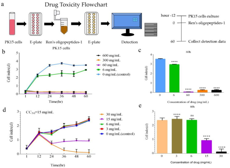
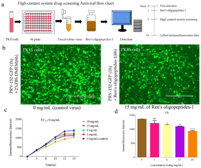
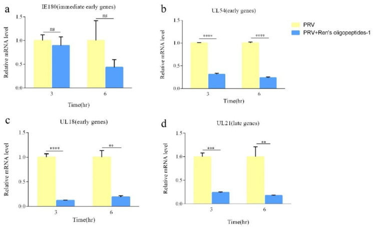
Newly synthesized Ren's oligopeptides-1 was found to have an antiviral effect in clinical trials, and the purpose of this study was to further demonstrate the antiviral activity of Ren's oligopeptides-1 against the PRV 152-GFP strain. We used the real-time cell analysis system (RTCA) to detect the cytotoxicity of different concentrations of Ren's oligopeptides-1. We then applied high content screening (HCS) to detect the antiviral activity of Ren's oligopeptides-1 against PRV. Meanwhile, the fluorescence signal of the virus was collected in real time and the expression levels of the related genes in the PK15 cells infected with PRV were detected using real-time PCR. At the mRNA level, we discovered that, at a concentration of 6 mg/mL, Ren's oligopeptides-1 reduced the expression of pseudorabies virus (PRV) genes such as IE180, UL18, UL54, and UL21 at a concentration of 6 mg/mL. We then determined that Ren's oligopeptides-1 has an EC50 value of 6 mg/mL, and at this level, no cytotoxicity was observed.

Keywords: RTCA; Ren’s oligopeptides-1; high content screening; pseudorabies virus (PRV).


Conflict of interest statement

The authors declare no conflict of interest.

Figures

Figure 1
Figure 1
Drug preparation flow chart of Ren’s oligopeptides-1.
Figure 2
Figure 2
Detection of the cytotoxicity of Ren’s oligopeptides-1 at different concentrations. (a) Flow chart of drug cytotoxicity assay using the real-time cell analysis (RTCA) method. (b) Cell index plots for 6 mg/mL, 60 mg/mL, 300 mg/mL, and 600 mg/mL drug concentrations. (c) Significant differences in the cell index of each group at 6 mg/mL, 60 mg/mL, 300 mg/mL, and 600 mg/mL drug concentrations at the 60 h. (d) Cell index plots for drug concentrations of 3 mg/mL, 6 mg/mL, 15 mg/mL, and 30 mg/mL. (e) Significant differences in the cell index of each group at the 60 h for drug concentrations of 3 mg/mL, 6 mg/mL, 15 mg/mL, and 30 mg/mL. ns, not significant (p > 0.05) and **** p < 0.0001 from the Student’s t-test. Error bars represent the SD from three independent experiments.
Figure 3
Figure 3
Ren’s oligopeptides-1 inhibits PRV-associated replication in PK15 cells. (a) High-content system drug screening antiviral flow chart. (b) Infected cells’ immunofluorescence plots were treated with 0 mg/mL and 15 mg/mL of Ren’s oligopeptides-1. (c) Infected cells were treated with 3 mg/mL, 6 mg/mL, 15 mg/mL, and 30 mg/mL of Ren’s oligopeptides-1; untreated and infected cells (PK15). A nonlinear regression analysis utilizing these data determined the EC50 for Ren’s oligopeptides-1 to be 6 mg/mL. (d) Significant differences in the immunofluorescence signal of each group at the 12 h for 3 mg/mL, 6 mg/mL, 15 mg/mL, and 30 mg/mL of Ren’s oligopeptides-1. ns, not significant (p > 0.05), * p < 0.05, ** p < 0.01, and **** p < 0.0001 from the Student’s t-test. Error bars represent the SD from five independent experiments.
Figure 4
Figure 4
Viral RNA expression as measured by real-time PCR in PRV-infected PK15 cells treated with 6 mg/mL of Ren’s oligopeptides-1. (a) Expression levels of IE180 at 3 and 6 h following treatment with Ren’s oligopeptides-1. (b) Expression levels of UL54 at 3 and 6 h following treatment with Ren’s oligopeptides-1. (c) Expression levels of UL18 at 3 and 6 h following treatment with Ren’s oligopeptides-1. (d) Expression levels of UL21 at 3 and 6 h following treatment with Ren’s oligopeptides-1. ns, not significant (p > 0.05); ** p < 0.01, *** p < 0.001, and **** p < 0.0001 from the Student’s t-test. Error bars represent SD from three independent experiments.

Similar articles

References

    1. Mettenleiter T.C. Aujeszky’s Disease and the Development of the Marker/DIVA Vaccination Concept. Pathogens. 2020;9:563. doi: 10.3390/pathogens9070563. - DOI - PMC - PubMed
    1. Karasneh G.A., Shukla D. Herpes simplex virus infects most cell types in vitro: Clues to its success. Virol. J. 2011;8:481. doi: 10.1186/1743-422X-8-481. - DOI - PMC - PubMed
    1. Ou J., Cai S., Zheng F., Lu G., Zhang G. Human pseudorabies virus infection: A new threat in China. J. Infect. 2019;80:578–606. doi: 10.1016/j.jinf.2019.12.018. - DOI - PubMed
    1. Ferrara G., Longobardi C., D’Ambrosi F., Amoroso M.G., D’Alessio N., Damiano S., Ciarcia R., Iovane V., Iovane G., Pagnini U., et al. Aujeszky’s Disease in South-Italian Wild Boars (Sus scrofa): A Serological Survey. Animals. 2021;11:3298. doi: 10.3390/ani11113298. - DOI - PMC - PubMed
    1. Tan L., Yao J., Yang Y., Luo W., Yuan X., Yang L., Wang A. Current Status and Challenge of Pseudorabies Virus Infection in China. Virol. Sin. 2021;36:588–607. doi: 10.1007/s12250-020-00340-0. - DOI - PMC - PubMed

LinkOut - more resources