

Post-Mortem Computed Tomography Pulmonary Findings in Harbor Porpoises (Phocoena phocoena)
- PMID: 35681918
- PMCID: PMC9179349
- DOI: 10.3390/ani12111454
Post-Mortem Computed Tomography Pulmonary Findings in Harbor Porpoises (Phocoena phocoena)
Abstract
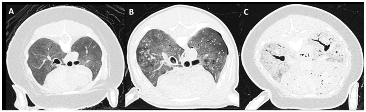
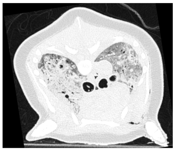
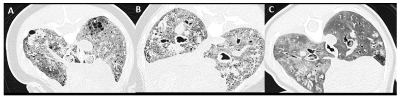
The application of whole-body post-mortem computed tomography (PMCT) in veterinary and wildlife post-mortem research programs is advancing. A high incidence of pulmonary pathology is reported in the harbor porpoise (Phocoena phocoena). In this study, the value of PMCT focused on pulmonary assessment is evaluated. The objectives of this study were to describe pulmonary changes as well as autolytic features detected by PMCT examination and to compare those findings with conventional necropsy. Retrospective evaluation of whole-body PMCT images of 46 relatively fresh harbor porpoises and corresponding conventional necropsy reports was carried out, with a special focus on the respiratory tract. Common pulmonary PMCT findings included: moderate (24/46) to severe (19/46) increased pulmonary soft tissue attenuation, severe parasite burden (17/46), bronchial wall thickening (30/46), and mild autolysis (26/46). Compared to conventional necropsy, PMCT more frequently identified pneumothorax (5/46 vs. none), tracheal content (26/46 vs. 7/46), and macroscopic pulmonary mineralization (23/46 vs. 11/46), and provided more information of the distribution of pulmonary changes. These results indicate that PMCT adds information on pulmonary assessment and is a promising complementary technique for necropsy, despite the frequent presence of mild autolytic features.
Keywords: cetacean; decomposition; necropsy; pulmonary pathology; virtopsy.
Conflict of interest statement
The authors declare no conflict of interest.
Figures


Similar articles
-
Fatal upper aerodigestive tract obstruction in an East Asian finless porpoise (Neophocaena asiaeorientalis sunameri): findings in post-mortem computed tomography.Forensic Sci Med Pathol. 2024 Jun;20(2):644-651. doi: 10.1007/s12024-023-00732-0. Epub 2023 Oct 13. Forensic Sci Med Pathol. 2024. PMID: 37831312
-
Characterisation of Crassicauda fuelleborni nematode infection in Indo-Pacific finless porpoises (Neophocaena phocaenoides) using postmortem computed tomography.Int J Parasitol Parasites Wildl. 2022 Apr 19;18:68-75. doi: 10.1016/j.ijppaw.2022.04.005. eCollection 2022 Aug. Int J Parasitol Parasites Wildl. 2022. PMID: 35519505 Free PMC article.
-
Illustrated cross-sectional computed tomography of the cetacean abdomino-pelvic organs.Ann Anat. 2024 Oct;256:152317. doi: 10.1016/j.aanat.2024.152317. Epub 2024 Aug 31. Ann Anat. 2024. PMID: 39182587
-
State of the art in post-mortem computed tomography: a review of current literature.Virchows Arch. 2019 Aug;475(2):139-150. doi: 10.1007/s00428-019-02562-4. Epub 2019 Apr 1. Virchows Arch. 2019. PMID: 30937612 Review.
-
Post-mortem CT imaging of the lungs: pathological versus non-pathological findings.Radiol Med. 2017 Dec;122(12):902-908. doi: 10.1007/s11547-017-0802-2. Epub 2017 Aug 23. Radiol Med. 2017. PMID: 28836139 Review.
References
-
- IJsseldijk L.L., Doeschate M.T.I.t., Davison N.J., Gröne A., Brownlow A.C. Crossing boundaries for cetacean conservation: Setting research priorities to guide management of harbour porpoises. Mar. Policy. 2018;95:77–84. doi: 10.1016/j.marpol.2018.07.006. - DOI
-
- Peltier H., Baagøe H.J., Camphuysen K.C., Czeck R., Dabin W., Daniel P., Deaville R., Haelters J., Jauniaux T., Jensen L.F., et al. The Stranding Anomaly as Population Indicator: The Case of Harbour Porpoise Phocoena phocoena in North-Western Europe. PLoS ONE. 2013;8:e62180. doi: 10.1371/journal.pone.0062180. - DOI - PMC - PubMed
-
- Van Bressem M., Raga J.A., Di Guardo G., Jepson P.D., Duignan P.J., Siebert U., Barrett T., de Oliveira Santos M.C., Moreno I.B., Siciliano S., et al. Emerging infectious diseases in cetaceans worldwide and the possible role of environmental stressors. Dis. Aquat. Org. 2009;86:143–157. doi: 10.3354/dao02101. - DOI - PubMed
-
- Hamel P.E.S., Giglio R.F., Cassle S.E., Farina L.L., Leone A.M., Walsh M.T. Postmortem computed tomography and magnetic resonance imaging findings in a case of coinfection of dolphin morbillivirus and aspergillus fumigatus in a juvenile bottlenose dolphin (Tursiops truncatus) J. Zoo Wildl. Med. 2020;51:448–454. doi: 10.1638/2019-0087. - DOI - PubMed
-
- Camphuysen C.J., Smeenk C., Addink M.J., Jansen O.E. Cetaceans stranded in the Netherlands from 1998 to 2007. Lutra. 2008;51:87–122.
Grants and funding
LinkOut - more resources
Full Text Sources
