. 2022 May 27;23(11):6039.
doi: 10.3390/ijms23116039.

Altered Brain Arginine Metabolism and Polyamine System in a P301S Tauopathy Mouse Model: A Time-Course Study

Affiliations

Altered Brain Arginine Metabolism and Polyamine System in a P301S Tauopathy Mouse Model: A Time-Course Study

Hannah Mein et al. Int J Mol Sci. .

Abstract

Altered arginine metabolism (including the polyamine system) has recently been implicated in the pathogenesis of tauopathies, characterised by hyperphosphorylated and aggregated microtubule-associated protein tau (MAPT) accumulation in the brain. The present study, for the first time, systematically determined the time-course of arginine metabolism changes in the MAPT P301S (PS19) mouse brain at 2, 4, 6, 8 and 12 months of age. The polyamines putrescine, spermidine and spermine are critically involved in microtubule assembly and stabilization. This study, therefore, further investigated how polyamine biosynthetic and catabolic enzymes changed in PS19 mice. There were general age-dependent increases of L-arginine, L-ornithine, putrescine and spermidine in the PS19 brain (particularly in the hippocampus and parahippocampal region). While this profile change clearly indicates a shift of arginine metabolism to favor polyamine production (a polyamine stress response), spermine levels were decreased or unchanged due to the upregulation of polyamine retro-conversion pathways. Our results further implicate altered arginine metabolism (particularly the polyamine system) in the pathogenesis of tauopathies. Given the role of the polyamines in microtubule assembly and stabilization, future research is required to understand the functional significance of the polyamine stress response and explore the preventive and/or therapeutic opportunities for tauopathies by targeting the polyamine system.

Keywords: PS19 mice; arginine; hippocampus; polyamine; polyamine stress response; spermine; tau; tauopathy.


Conflict of interest statement

The authors declare no conflict of interest. The funders had no role in the design of the study; in the collection, analyses, or interpretation of data, in the writing of the manuscript or in the decision to publish the results.

Figures
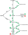
Figure 1
Figure 1
L-arginine metabolic pathways and the polyamine system. L-arginine is metabolized by nitric oxide synthase (NOS), arginase and arginine decarboxylase (ADC) to form several bioactive molecules and can be de novo synthesized by argininosuccinate synthetase (ASS) and argininosuccinate lyase (ASL) from L-citrulline (see text for detailed description). Overall, we found genotype- and age-related increases in L-arginine, L-ornithine, glutamine, putrescine and spermidine (indicated by short green arrows) and decreases in glutamate and spermine (indicated by short red arrows) levels in the brain of PS19 mice, indicating a shift of L-arginine metabolism towards the arginase and polyamine system. Moreover, PS19 mice displayed increased levels of mRNA and/or protein expression of ASS, ASL, arginase I and II, ornithine decarboxylase (ODC), spermidine synthase (SPDS), spermine oxidase (SMOX), spermidine/spermine-N1-acetyltransferase-1 (SSAT1) and polyamine oxidase (PAO), suggesting upregulated citrulline recycling and polyamine retro-conversion (indicated by long green arrows). AGMAT, agmatinase; SMS, spermine synthase. Adapted from [28].
Figure 2
Figure 2
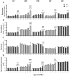
Mean (± SEM) levels of L-arginine (A) and its three direct metabolites L-citrulline (B), L-ornithine (C) and agmatine (D) in the frontal cortex (FC), hippocampus (HP), parahippocampal region (PH), striatum (ST) and cerebellum (CE) of wildtype (WT) and PS19 mice at 2, 4, 6, 8 and 12 months of age (n = 7–9/genotype/age). & indicates a significant genotype and age interaction at & p < 0.05, && p < 0.01 or &&& p < 0.001. # indicates a significant age effect at # p < 0.05, ## p < 0.01, ### p < 0.001 or #### p < 0.0001. * indicates a significant genotype effect at * p < 0.05, ** p < 0.01 or **** p < 0.0001.
Figure 3
Figure 3
Mean (± SEM) levels of glutamine (A), glutamate (B), the glutamine/glutamate ratio (C) and GABA (D) in the frontal cortex (FC), hippocampus (HP), parahippocampal region (PH), striatum (ST) and cerebellum (CE) of wildtype (WT) and PS19 mice at 2, 4, 6, 8 and 12 months of age (n = 7–9/genotype/age). & indicates a significant genotype and age interaction at & p < 0.05, && p < 0.01 or &&& p < 0.001. # indicates a significant age effect at ## p < 0.01, ### p < 0.001 or #### p < 0.0001. * indicates a significant genotype effect at * p < 0.05, ** p < 0.01, *** p < 0.001 or **** p < 0.0001.
Figure 4
Figure 4
Mean (± SEM) levels of putrescine (A), spermidine (B), spermine (C) and the spermidine/spermine ratio (D) in the frontal cortex (FC), hippocampus (HP), parahippocampal region (PH), striatum (ST) and cerebellum (CE) of wildtype (WT) and PS19 mice at 2, 4, 6, 8 and 12 months of age (n = 7–9/genotype/age). & indicates a significant genotype and age interaction at & p < 0.05, &&& p < 0.001 or &&&& p < 0.0001. # indicates a significant age effect at # p < 0.05, ### p < 0.001 or ####p < 0.0001. * indicates a significant genotype effect at * p < 0.05, ** p < 0.01, *** p < 0.001 or **** p < 0.0001.
Figure 5
Figure 5
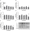
Mean (± SEM) relative fold change (2−ΔΔCt) of mRNA expression of arginase I (A), arginase II (B), ornithine decarboxylase (ODC, C), arginine decarboxylase (ADC, D), agmatinase (AGMAT, E), spermidine synthase (SPDS, F), spermine synthase (SMS, G), spermine oxidase (SMOX, H), spermidine/spermine-N1-acetyltransferase-1 (SSAT1, I) and polyamine oxidase (PAO, J) in the frontal cortex (FC), hippocampus (HP), parahippocampal region (PH) and cerebellum (CE) of wildtype (WT) and PS19 mice at 8 and 12 months (m) of age (n = 6–8/genotype/age). & indicates a significant genotype and age interaction at & p < 0.05 or && p < 0.01. # indicates a significant age effect at # p < 0.05 or ## p < 0.01. * indicates a significant genotype effect at * p < 0.05, ** p < 0.01, *** p < 0.001 or **** p < 0.0001.
Figure 6
Figure 6
Mean (± SEM) protein expression of arginase I (AI; A), arginase II (AII; B), ornithine decarboxylase (ODC, C), argininosuccinate synthetase (ASS, D) and argininosuccinate lyase (ASL, E) in the parahippocampal region of wildtype (WT) and PS19 mice at 2, 4, 6, 8 and 12 months of age (n = 7–9/genotype/age). (F): representative western blots of AI, AII, ODC, ASS and ASL, as well as housekeeping protein glyceraldehyde 3-phosphate dehydrogenase (GAPDH), in wild-type (W) and PS19 (P) mice at five age points. && indicates a significant genotype and age interaction at p < 0.01. # indicates a significant age effect at # p < 0.05 or ## p < 0.01. * indicates a significant genotype effect at ** p < 0.01 or *** p < 0.001.
Figure 7
Figure 7
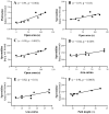
Scattergrams showing the significant correlations between the time spent in the open arms of the elevated plus-maze test and putrescine in the frontal cortex (A), spermidine in the frontal cortex (B) and parahippocampal region (C), between the total number of arm entries in the elevated plus-maze and spermidine in the hippocampus (D) and parahippocampal region (E) and between the total path length generated in the open field test and spermidine in the hippocampus (F), in 8-month-old PS19 mice.

Similar articles

Cited by

References

    1. Arendt T., Stieler J.T., Holzer M. Tau and Tauopathies. Brain Res. Bull. 2016;126:238–292. doi: 10.1016/j.brainresbull.2016.08.018. - DOI - PubMed
    1. Wang Y., Mandelkow E. Tau in Physiology and Pathology. Nat. Rev. Neurosci. 2016;17:5–21. doi: 10.1038/nrn.2015.1. - DOI - PubMed
    1. Weingarten M.D., Lockwood A.H., Hwo S.Y., Kirschner M.W. A Protein Factor Essential for Microtubule Assembly. Proc. Natl. Acad. Sci. USA. 1975;72:1858–1862. doi: 10.1073/pnas.72.5.1858. - DOI - PMC - PubMed
    1. Kovacs G.G. Tauopathies. Handb. Clin. Neurol. 2017;145:355–368. doi: 10.1016/b978-0-12-802395-2.00025-0. - DOI - PubMed
    1. Lebouvier T., Pasquier F., Buée L. Update on Tauopathies. Curr. Opin. Neurol. 2017;30:589–598. doi: 10.1097/WCO.0000000000000502. - DOI - PubMed

LinkOut - more resources