. 2022 May 29;23(11):6092.
doi: 10.3390/ijms23116092.

Chemogenetic Activation of Astrocytes in the Basolateral Amygdala Contributes to Fear Memory Formation by Modulating the Amygdala-Prefrontal Cortex Communication

Affiliations

Chemogenetic Activation of Astrocytes in the Basolateral Amygdala Contributes to Fear Memory Formation by Modulating the Amygdala-Prefrontal Cortex Communication

Zhuogui Lei et al. Int J Mol Sci. .

Abstract

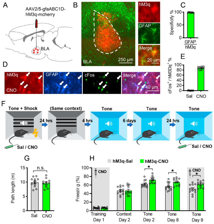
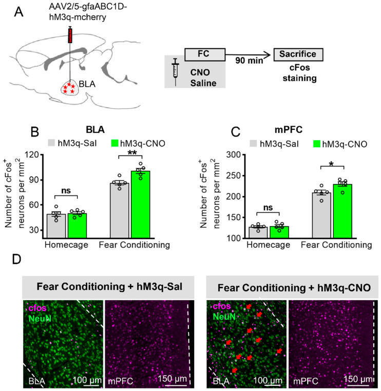
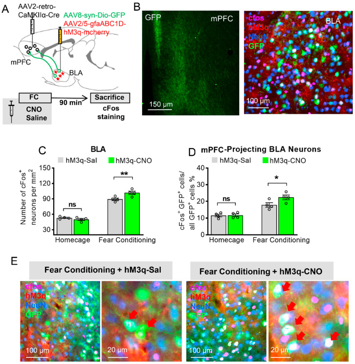
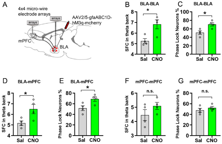
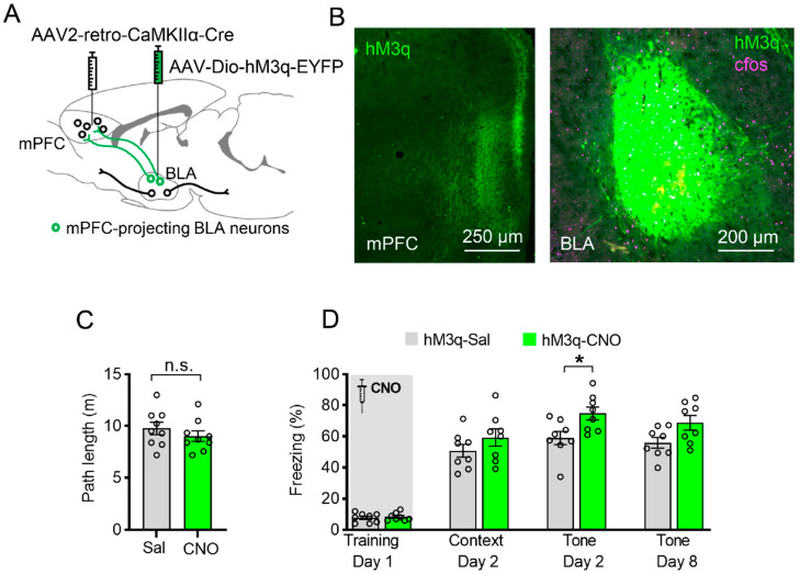
The basolateral amygdala (BLA) is one of the key brain areas involved in aversive learning, especially fear memory formation. Studies of aversive learning in the BLA have largely focused on neuronal function, while the role of BLA astrocytes in aversive learning remains largely unknown. In this study, we manipulated the BLA astrocytes by expressing the Gq-coupled receptor hM3q and discovered that astrocytic Gq modulation during fear conditioning promoted auditorily cued fear memory but did not affect less stressful memory tasks or induce anxiety-like behavior. Moreover, chemogenetic activation of BLA astrocytes during memory retrieval had no effect on fear memory expression. In addition, astrocytic Gq activation increased c-Fos expression in the BLA and the medial prefrontal cortex (mPFC) during fear conditioning, but not in the home cage. Combining these results with retrograde virus tracing, we found that the activity of mPFC-projecting BLA neurons showed significant enhancement after astrocytic Gq activation during fear conditioning. Electrophysiology recordings showed that activating astrocytic Gq in the BLA promoted spike-field coherence and phase locking percentage, not only within the BLA but also between the BLA and the mPFC. Finally, direct chemogenetic activation of mPFC-projecting BLA neurons during fear conditioning enhanced cued fear memory. Taken together, our data suggest that astrocytes in the BLA may contribute to aversive learning by modulating amygdala-mPFC communication.

Keywords: anxiety; astrocytes; basolateral amygdala; chemogenetic; electrophysiology; fear conditioning; medial prefrontal cortex; neurons; projection.


Conflict of interest statement

The authors declare no conflict of interest.

Figures

Figure 1
Figure 1
Astrocytic Gq activation in the BLA promotes auditorily cued fear memory formation. (A) Schematic picture showing AAV-gfaABC1D-hM3q-mcherry injection in the BLA bilaterally. (B) hM3q virus expression in the BLA (left panel); red is hM3q, green is GFAP staining, scale bar 250 µm. Magnified view of hM3q co-expressed with GFAP (right panel); scale bar 20 µm. (C) Cells with hM3q expression are highly co-labeled with GFAP (98.33 ± 0.5%, n = 3, 9 slices). (D,E) CNO administration to mice expressing hM3q (red) in BLA astrocytes resulted in a significant increase in c-Fos expression (green) in these astrocytes (white arrow), compared to saline-injected controls (p < 0.0001, n = 3 per groups, 9 slices per group; blue is GFAP staining, scale bar 40μm). (F) Schematic diagram of fear memory training and contextual/auditorily cued fear memory retrieval. (G) Path length of exploration in the conditioning cage after CNO or saline injection (p > 0.05, unpaired Student’s t-test, n.s. stand for no significance). (H) Chemogenetic activation of BLA astrocytes increased auditorily cued fear memory on day 2 (p = 0.0367), day 8 (p = 0.0424), but not contextual fear memory (p > 0.05) compared to saline group. On day 9, CNO injection before auditorily cued memory test still showed significant enhancement of fear memory compared to saline group (p = 0.0361) (Saline group n = 12, CNO group n = 11, * stands for p < 0.05, two-way ANOVA followed by Bonferroni’s post hoc test, main effect of Sal/CNO: F (1, 21) = 7.530, p = 0.0122). Data are presented as mean ± SEM.
Figure 2
Figure 2
Astrocytic Gq activation increases c-Fos expression in the BLA and the mPFC in fear conditioning, but not in the home cage. (A) Schematic diagram for brain sample collection and c-Fos staining from mice injected with AAV2/5-gfaABC1D-hM3q-mcherry. (B) c-Fos level in BLA neurons of four-group mice expressing hM3q in their BLA astrocytes (n = 5 of each group, 5 slices for each mouse, two-way ANOVA with Bonferroni post hoc comparison test, main effect of Sal/CNO: F (1, 8) = 6.461, p = 0.0346, ‘homecage (HC) + hM3q-Sal’ versus ‘HC + hM3q-CNO’: p > 0.9999, ns stands for no significance, ‘fear conditioning (FC) + hM3q-Sal’ versus ‘FC + hM3q-CNO’: p = 0.0027, ** stands for p < 0.01, ns stands for no significance). (C) c-Fos level in mPFC neurons of four-group mice expressing hM3q in their BLA astrocytes (n = 5 of each group, two-way ANOVA with Bonferroni post hoc comparison test, main effect of Sal/CNO: F (1, 8) = 8.189, p = 0.0211, ‘HC + hM3q-Sal’ versus ‘HC + hM3q-CNO’: p > 0.9999, ns stands for no significance, ‘FC + hM3q-Sal’ versus ‘FC + hM3q-CNO’: p = 0.0127, * stands for p < 0.05). (D) Representative pictures of c-Fos (pink) expression in BLA neurons (NeuN, green) and mPFC under CNO or saline after fear conditioning, scale bar 100 μm, 150 μm. Red arrow shows the BLA neurons (NeuN) with c-Fos expression. All data are presented as mean ± SEM.
Figure 3
Figure 3
Astrocytic Gq modulation induces a projection-specific enhancement of BLA–mPFC neurons during fear learning. (A) Schematic diagram of targeting mPFC-projecting BLA neurons: AAV2-retro-CamkII-Cre was injected into the mPFC, and AAV8-syn-Dio–GFP together with AAV2/5-gfaABC1D-hM3q-mcherry was injected into the BLA. (B) Left: GFP-positive axons of BLA projection neurons are clearly visible in the mPFC. Right: mPFC-projecting BLA neurons (green) with c-Fos (pink) and NeuN (blue) staining, and hM3q (red) in BLA astrocytes. (C) c-Fos level in BLA neurons of four-group mice expressing GFP in mPFC-projecting BLA neurons and hM3q in their BLA astrocytes (n = 4 of each group, 5 slices for each mouse, two-way ANOVA with Bonferroni post hoc comparison test, main effect of Sal/CNO: F (1, 6) = 6.423, p = 0.0444, ‘homecage (HC) + hM3q-Sal’ versus ‘HC + hM3q-CNO’: p > 0.9999, ‘fear conditioning (FC) + hM3q-Sal’ versus ‘FC + hM3q-CNO’: p = 0.0055, ** stands for p < 0.01, ns stands for no significance). (D) Fear-conditioned mice injected with CNO showed increase in the percentage of mPFC-projecting BLA neurons that express c-Fos compared with FC mice injected with saline (n = 4 of each group, two-way ANOVA with Bonferroni post hoc comparison test, main effect of Sal/CNO: F (1, 6) = 7.424, p = 0.0344, ‘HC + hM3q-Sal’ versus ‘HC + hM3q-CNO’: p > 0.9999, ‘FC + hM3q-Sal’ versus ‘FC + hM3q-CNO’: p = 0.032, * stands for p < 0.05, ns stands for no significance). (E) Representative images of hM3q (red), c-Fos (pink), mPFC-projecting BLA neurons (green), and NeuN (Blue) in the BLA; red arrow shows the mPFC-projecting BLA neurons with c-Fos expression. All data are presented as mean ± SEM.
Figure 4
Figure 4
Electrophysiology recording showing that activation of astrocytic Gq in the BLA promotes spike-field coherence and phase locking within the BLA and between the BLA and the mPFC. (A) Schematic of the double-recording electrodes implanted in the BLA and the mPFC. (B,C) Quantification of spike-field coherence (SFC) within BLA in theta band and phase locking percentage within BLA between CNO and saline treatment (n = 4 per group, neurons number = 24, p = 0.0165, p = 0.0161, by unpaired Student’s t-test). (D,E) SFC between BLA and mPFC in theta band and phase locking percentage between BLA and mPFC with CNO treatment was increased compared with saline group (n = 4 per group, neurons number = 24, p = 0.0360, p = 0.0336, by unpaired Student’s t-test). (F,G) CNO application had no significant effect on SFC and phase locking neuron percentage within mPFC (n = 4 per group, neurons number = 24, p = 0.2066, p = 0.3688, by unpaired Student’s t-test). * stands for p < 0.05, n.s. stands for no significance All data are presented as mean ± SEM.
Figure 5
Figure 5
Specific chemogenetic activation of mPFC-projecting BLA neurons during learning enhances auditorily cued fear memory. (A) Schematic of the experiment: AAV2-retro-CaMKIIα-Cre was injected into the mPFC, and AAV2/9-syn-DIO–hM3q–EYFP was injected into the BLA. (B) Left: hM3q-positive axons (green) of BLA projection neurons are clearly visible in the mPFC. Right: hM3q (green) in BLA neurons projecting to the mPFC with c-Fos staining (pink). (C) Mice expressing hM3q in the mPFC-projecting BLA neurons were injected with either saline or CNO 30 min before FC training. CNO application before training had no significant effect on motor behavior (p = 0.3531, unpaired Student’s t-test, n.s. stands for no significance). (D) CNO application had no significant effect on contextual freezing level on day 2 (p > 0.4811) but promoted auditorily cued fear memory on day 2 (p = 0.0217) (Saline group n = 8, CNO group n = 8, * stands for p < 0.05, two-way ANOVA followed by Bonferroni’s post hoc test, main effect of Sal/CNO: F (1, 14) = 6.968, p = 0.0194). All data are presented as mean ± SEM.

Similar articles

Cited by

References

    1. Letzkus J.J., Wolff S.B.E., Meyer E.M.M., Tovote P., Courtin J., Herry C., Lüthi A. A Disinhibitory Microcircuit for Associative Fear Learning in the Auditory Cortex. Nature. 2011;480:331–335. doi: 10.1038/nature10674. - DOI - PubMed
    1. Aggleton J.P. The Amygdala: A Functional Analysis. Oxford University Press; Oxford, UK: 2000.
    1. Bocchio M., Nabavi S., Capogna M. Synaptic Plasticity, Engrams, and Network Oscillations in Amygdala Circuits for Storage and Retrieval of Emotional Memories. Neuron. 2017;94:731–743. doi: 10.1016/j.neuron.2017.03.022. - DOI - PubMed
    1. Herry C., Johansen J.P. Encoding of Fear Learning and Memory in Distributed Neuronal Circuits. Nat. Neurosci. 2014;17:1644–1654. doi: 10.1038/nn.3869. - DOI - PubMed
    1. Maren S. Synaptic Mechanisms of Associative Memory in the Amygdala. Neuron. 2005;47:783–786. doi: 10.1016/j.neuron.2005.08.009. - DOI - PubMed

LinkOut - more resources