. 2022 May 29;23(11):6104.
doi: 10.3390/ijms23116104.

Sophoraflavanone G from Sophora flavescens Ameliorates Allergic Airway Inflammation by Suppressing Th2 Response and Oxidative Stress in a Murine Asthma Model

Affiliations

Sophoraflavanone G from Sophora flavescens Ameliorates Allergic Airway Inflammation by Suppressing Th2 Response and Oxidative Stress in a Murine Asthma Model

Meng-Chun Wang et al. Int J Mol Sci. .

Abstract

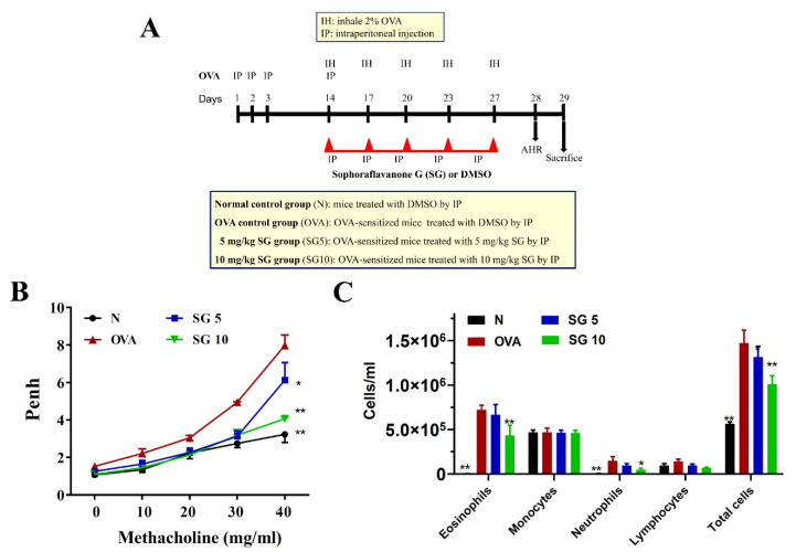
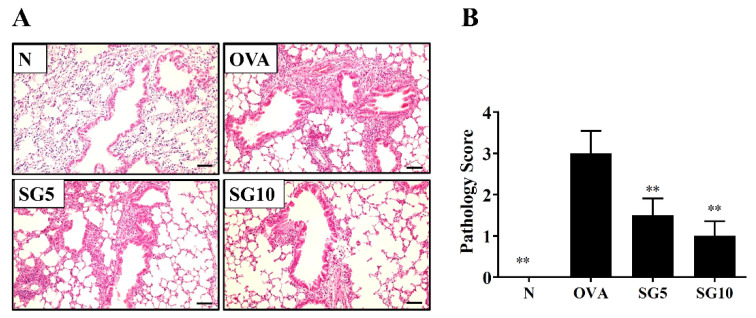
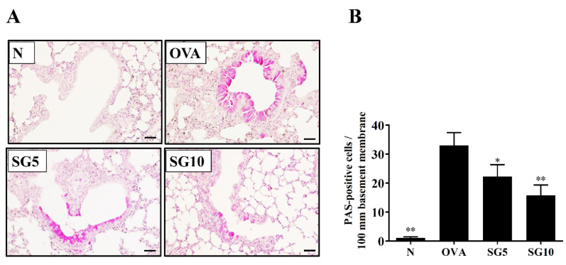
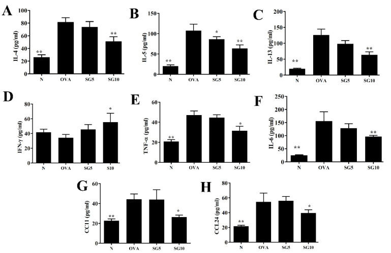
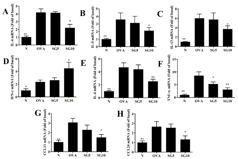
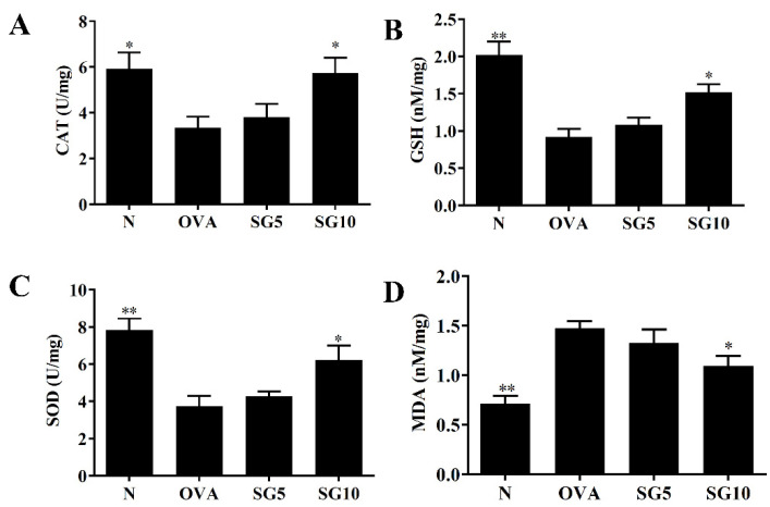
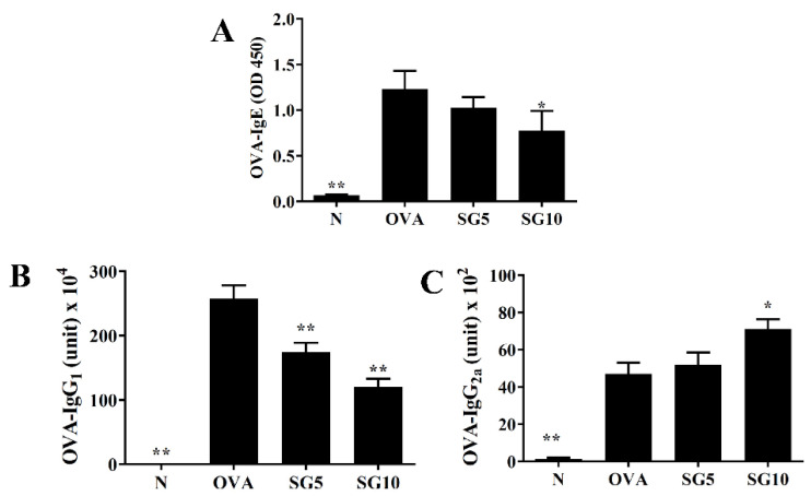
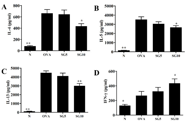
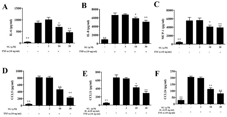
Sophoraflavanone G (SG), isolated from Sophora flavescens, has anti-inflammatory and anti-tumor bioactive properties. We previously showed that SG promotes apoptosis in human breast cancer cells and leukemia cells and reduces the inflammatory response in lipopolysaccharide-stimulated macrophages. We investigated whether SG attenuates airway hyper-responsiveness (AHR) and airway inflammation in asthmatic mice. We also assessed its effects on the anti-inflammatory response in human tracheal epithelial cells. Female BALB/c mice were sensitized with ovalbumin, and asthmatic mice were treated with SG by intraperitoneal injection. We also exposed human bronchial epithelial BEAS-2B cells to different concentrations of SG to evaluate its effects on inflammatory cytokine levels. SG treatment significantly reduced AHR, eosinophil infiltration, goblet cell hyperplasia, and airway inflammation in the lungs of asthmatic mice. In the lungs of ovalbumin-sensitized mice, SG significantly promoted superoxide dismutase and glutathione expression and attenuated malondialdehyde levels. SG also suppressed levels of Th2 cytokines and chemokines in lung and bronchoalveolar lavage samples. In addition, we confirmed that SG decreased pro-inflammatory cytokine, chemokine, and eotaxin expression in inflammatory BEAS-2B cells. Taken together, our data demonstrate that SG shows potential as an immunomodulator that can improve asthma symptoms by decreasing airway-inflammation-related oxidative stress.

Keywords: Th2 cell; airway hyper-responsiveness; airway inflammation; asthma; sophoraflavanone G.


Conflict of interest statement

The authors have no conflict of interest to declare.

Figures

Figure 1
Figure 1
Effect of sophoraflavanone G (SG) on pulmonary function in asthmatic mice. (A) Experimental procedures for asthmatic mouse studies. (B) Enhanced pauses (Penh) were detected by methacholine inhalation (0–40 mg/mL) in mice. (C) Numbers of inflammatory and total cells in BALF. The data are presented as mean ± SEM of three independent experiments (n = 10 per group); * p < 0.05, ** p < 0.01 versus OVA group.
Figure 2
Figure 2
Sophoraflavanone G (SG) reduces eosinophil infiltration in the mouse lung. (A) H&E staining showing eosinophil infiltration (200× magnification). (B) Inflammatory scores are for lung sections. The data are presented as mean ± SEM of three independent experiments (n = 4–6 per group); ** p < 0.01 versus the OVA group (scale bar = 100 µm).
Figure 3
Figure 3
Sophoraflavanone G (SG) reduces goblet cell hyperplasia in the mouse lung. (A) PAS staining showing goblet cell hyperplasia (200× magnification). (B) PAS-positive cells were calculated per 100 μm in the trachea. The data are presented as mean ± SEM of three independent experiments (n = 4–6 per group); * p < 0.05, ** p < 0.01 versus the OVA group (scale bar = 100 µm).
Figure 4
Figure 4
Sophoraflavanone G (SG) regulates BALF cytokine and chemokine levels. (A) IL-4, (B) IL-5, (C) IL-13, (D) IFN-γ, (E) TNF-α, (F) IL-6, (G) CCL11, and (H) CCL24 as measured by ELISA. The data are presented as mean ± SEM of three independent experiments (n = 10 per group); * p < 0.05, ** p < 0.01 versus the OVA group.
Figure 5
Figure 5
Sophoraflavanone G (SG) regulates cytokine and chemokine gene expression in the lung. (A) IL-4, (B) IL-5, (C) IL-13, (D) IFN-γ, (E) IL-6, (F) TNF-α, (G) CCL11, and (H) CCL24 were determined by real-time PCR. Fold values relative to β-actin expression. The data are presented as mean ± SEM of three independent experiments (n = 10 per group); * p < 0.05, ** p < 0.01 versus the OVA group.
Figure 6
Figure 6
Sophoraflavanone G (SG) regulates oxidative stress factors. (A) CAT, (B) GSH, (C) SOD, and (D) MDA were measured in lung tissues. The data are presented as mean ± SEM of three independent experiments (n = 10 per group); * p < 0.05, ** p < 0.01 versus the OVA group.
Figure 7
Figure 7
Sophoraflavanone G (SG) regulated OVA-specific antibodies in serum. (A) OVA-IgE, (B) OVA-IgG1, and (C) OVA-IgG2a were detected by ELISA. The data are presented as mean ± SEM of three independent experiments (n = 10 per group); * p < 0.05, ** p < 0.01 versus the OVA group.
Figure 8
Figure 8
Sophoraflavanone G (SG) regulates cytokine production in OVA-activated splenocytes. (A) IL-4, (B) IL-5, (C) IL-13, and (D) IFN-γ levels were examined by ELISA. The data are presented as mean ± SEM of three independent experiments (n = 10 per group); * p < 0.05, ** p < 0.01 versus the OVA group.
Figure 9
Figure 9
Sophoraflavanone G (SG) reduces inflammatory cytokine and chemokine production in BEAS-2B cells. BEAS-2B cells (6–8 passages) were treated with SG, then stimulated with TNF-α (10 ng/mL) for 24 h. (A) IL-6, (B) IL-8, (C) MCP-1, and (D) CCL5 were examined by ELISA. Values are mean ± SEM; * p < 0.05, ** p < 0.01 versus BEAS-2B cells stimulated with TNF-α alone. (E,F) Cells also were treated with SG and then stimulated with 10 ng/mL TNF-α/IL-4 for 24 h. (E) CCL11 and (F) CCL24 were detected. The data are presented as mean ± SEM of three independent experiments (n = 12 per group); * p < 0.05, ** p < 0.01 versus BEAS-2B cells stimulated with TNF-α /IL-4.

Similar articles

Cited by

References

    1. Papi A., Brightling C., Pedersen S.E., Reddel H.K. Asthma. Lancet. 2018;391:783–800. doi: 10.1016/S0140-6736(17)33311-1. - DOI - PubMed
    1. Castillo J.R., Peters S.P., Busse W.W. Asthma exacerbations: Pathogenesis, prevention, and treatment. J. Allergy Clin. Immunol. Pract. 2017;5:918–927. doi: 10.1016/j.jaip.2017.05.001. - DOI - PMC - PubMed
    1. Patel S.J., Teach S.J. Asthma. Pediatr. Rev. 2019;40:549–567. doi: 10.1542/pir.2018-0282. - DOI - PMC - PubMed
    1. Esteban-Gorgojo I., Antolín-Amérigo D., Domínguez-Ortega J., Quirce S. Non-eosinophilic asthma: Current perspectives. J. Asthma Allergy. 2018;11:267–281. doi: 10.2147/JAA.S153097. - DOI - PMC - PubMed
    1. Seys S.F., Lokwani R., Simpson J.L., Bullens D.M.A. New insights in neutrophilic asthma. Curr. Opin. Pulm. Med. 2019;25:113–120. doi: 10.1097/MCP.0000000000000543. - DOI - PubMed