. 2022 May 29;23(11):6107.
doi: 10.3390/ijms23116107.

Curvicollide D Isolated from the Fungus Amesia sp. Kills African Trypanosomes by Inhibiting Transcription

Affiliations

Curvicollide D Isolated from the Fungus Amesia sp. Kills African Trypanosomes by Inhibiting Transcription

Matilde Ortiz-Gonzalez et al. Int J Mol Sci. .

Abstract

Sleeping sickness or African trypanosomiasis is a serious health concern with an added socio-economic impact in sub-Saharan Africa due to direct infection in both humans and their domestic livestock. There is no vaccine available against African trypanosomes and its treatment relies only on chemotherapy. Although the current drugs are effective, most of them are far from the modern concept of a drug in terms of toxicity, specificity and therapeutic regime. In a search for new molecules with trypanocidal activity, a high throughput screening of 2000 microbial extracts was performed. Fractionation of one of these extracts, belonging to a culture of the fungus Amesia sp., yielded a new member of the curvicollide family that has been designated as curvicollide D. The new compound showed an inhibitory concentration 50 (IC50) 16-fold lower in Trypanosoma brucei than in human cells. Moreover, it induced cell cycle arrest and disruption of the nucleolar structure. Finally, we showed that curvicollide D binds to DNA and inhibits transcription in African trypanosomes, resulting in cell death. These results constitute the first report on the activity and mode of action of a member of the curvicollide family in T. brucei.

Keywords: African trypanosomiasis; curvicollide D; natural products; new trypanocidal molecule.


Conflict of interest statement

The authors declare no conflict of interest.

Figures

Figure 1
Figure 1
Structure of curvicollide D.
Figure 2
Figure 2
Key COSY and TOCSY correlations (bold bonds) determining the different spin systems of curvicollide D. Key HMBC correlations (blue arrows, H to C) connecting the different spin systems and rendering the connectivity of curvicollide D.
Figure 3
Figure 3
Key NOESY correlations (red dashed arrows) and coupling constants determining the relative stereochemistry of the stereoclusters C3–C4 and C9–C11 in curvicollide D.
Figure 4
Figure 4
Curvicollide D inhibits proliferation and affects the cell cycle of bloodstream form of T. brucei. Resazurin in vitro cytotoxicity assay was used to determine the curve dose response (viability percentage versus curvicollide D concentration). The IC50 was determined interpolation, using GraphPad software (A) IC50 of T. brucei and (B) IC50 of the human hepatocarcinoma cells (Hep G2) during 24 h. (C,D) Flow cytometry analysis of the cell cycle after 6 and 15 h of exposure to curvicollide D (1.5 μM) or after treatment with DMSO (control). Percentages for each phase were determined using FlowJo software. (PE) phycoerythrin. Data represent mean and standard deviation from three independent experiments. Statistical significance between control and treatmens at 6 and 15 h was estimated using Student’s two-tailed t-test. p-Values were expressed as follows: ** p < 0.01 and *** p < 0.001.
Figure 5
Figure 5
Changes in cell morphology and nucleolar structure after 3, 6 and 15 h of treatment with curvicollide D. Bloodstream forms of T. brucei incubated with 1.5 μM of Curvicollide D for 3, 6 and 15 h and cells treated with DMSO (control). (A) Percentage of abnormal cells. Error bars represent the standard deviations from three independent experiments. Statistical significance was *, p < 0.05; ***, p < 0.001 using ANOVA and Bonferroni post hoc test. (B) Changes in cell morphology after 3, 6 and 15 h of treatment. DAPI-stained parasites were observed by confocal microscopy (top line) and so was differential interference contrast (DIC) with DAPI (bottom line). The nucleus (N) and kinetoplast (K) are shown in white. The arrow in red indicates the nucleolus (n). (C) Percentage of cells that maintain the nucleolus structure after treatment. (D) Confocal microscopy showing disassembly of the nucleolus after a 3 h treatment with curvicollide D. (E) Immunofluorescence microscopy using an antibody against the nucleolar marker L1C6 (green) after a 3 h treatment with curvicollide D. Scale bar, 5 μm. Statistical significance between control and 3 h of treatment was estimated using Student’s two-tailed t-test. p-Value was expressed as follows: *** p < 0.001.
Figure 6
Figure 6
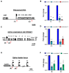
Inhibition of RNA Pol I and Pol II transcription by curvicollide D in bloodstream forms of T. brucei. (A) Schematic ribosomal DNA (rDNA) transcription unit in T. brucei, indicating the location of the primer sets (1) and (2). (C) The VSG221 expression site and (E) the splice leader locus with the promoters indicated as a black flag. Genes are shown with grey boxes. T. brucei 221 was treated with 1.5 μM of curvicollide D for 3 h. (B) Pol I RNA precursor transcripts were analysed in the Pol I transcribed rDNA region (primer pairs (1) and (2)) and so was (D) the active VSG221 ES (primer pairs (3) and (4)). (F) RNA pol II precursor transcripts were analysed in the splice leader locus using primer pairs (5) and (6). Actinomicin D and α-amanitin were used as positive controls of inhibition for RNA Pol I and Pol II transcription respectively, and cells treated with DMSO as a negative control. Error bars represent the standard deviation from three independent experiments. Statistical significance was **, p < 0.01; ***, p < 0.001 using ANOVA and Bonferroni post hoc test.
Figure 7
Figure 7
Curvicollide D intercalates into DNA. Ethidium bromide displacement assay using a double stranded DNA plasmid. Increasing concentration of curvicollide D (10 μM to 30 μM) were used to determine (A) the fluorescence intensity (arbitrary unit (a.u)) from 550 nm wavelength to 720 nm, and (B) the percentage of EtBr fluorescence at 610 nm determined in (A). Mean values of triplicates and corresponding standard deviations are shown. Statistical significance between control (EtBr) and each treatment was stimated using Student’s two-tailed t-test. p-Values were expressed as follows: * p < 0.05; ** p < 0.01 and *** p < 0.001.

Similar articles

Cited by

References

    1. Kennedy P.G.E., Rodgers J. Clinical and Neuropathogenetic Aspects of Human African Trypanosomiasis. Front. Immunol. 2019;10:39. doi: 10.3389/fimmu.2019.00039. - DOI - PMC - PubMed
    1. [(accessed on 25 April 2022)]. Available online: https://www.who.int/news-room/fact-sheets/detail/trypanosomiasis-human-a...
    1. Yaro M., Munyard K.A., Stear M.J., Groth D.M. Combatting African Animal Trypanosomiasis (AAT) in livestock: The potential role of trypanotolerance. Vet. Parasitol. 2016;225:43–52. doi: 10.1016/j.vetpar.2016.05.003. - DOI - PubMed
    1. Babokhov P., Sanyaolu A.O., Oyibo W.A., Fagbenro-Beyioku A.F., Iriemenam N.C. A current analysis of chemotherapy strategies for the treatment of human African trypanosomiasis. Pathog. Glob. Health. 2013;107:242–252. doi: 10.1179/2047773213Y.0000000105. - DOI - PMC - PubMed
    1. Garcia-Salcedo J.A., Unciti-Broceta J.D., Valverde-Pozo J., Soriano M. New Approaches to Overcome Transport Related Drug Resistance in Trypanosomatid Parasites. Front. Pharmacol. 2016;7:351. doi: 10.3389/fphar.2016.00351. - DOI - PMC - PubMed

Substances