

Interaction of Proteins with Inverted Repeats and Cruciform Structures in Nucleic Acids
- PMID: 35682854
- PMCID: PMC9180970
- DOI: 10.3390/ijms23116171
Interaction of Proteins with Inverted Repeats and Cruciform Structures in Nucleic Acids
Abstract
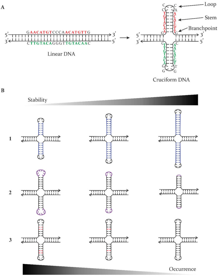
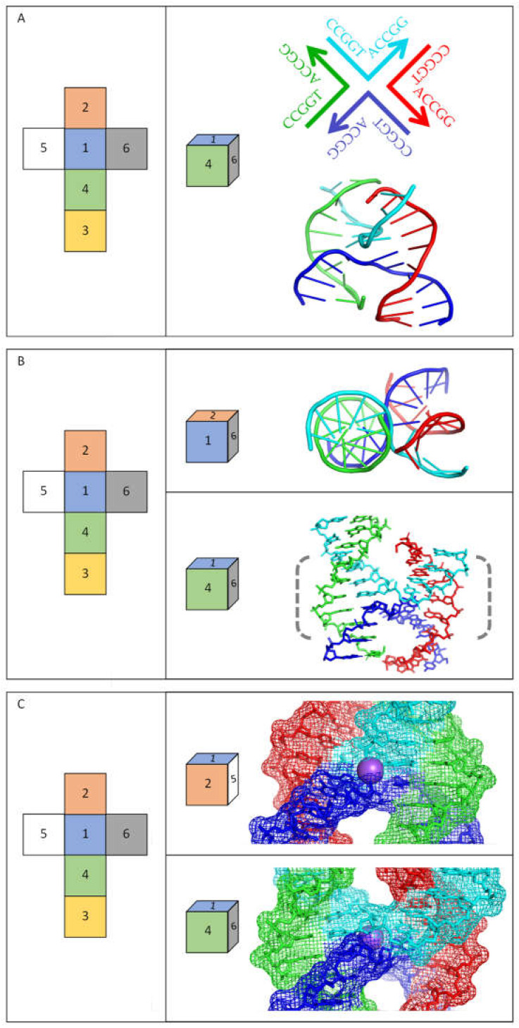
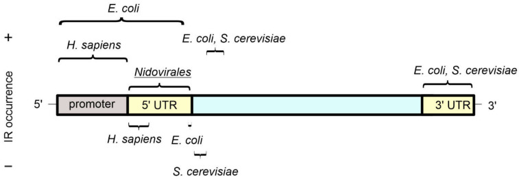
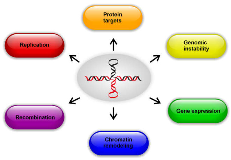
Cruciforms occur when inverted repeat sequences in double-stranded DNA adopt intra-strand hairpins on opposing strands. Biophysical and molecular studies of these structures confirm their characterization as four-way junctions and have demonstrated that several factors influence their stability, including overall chromatin structure and DNA supercoiling. Here, we review our understanding of processes that influence the formation and stability of cruciforms in genomes, covering the range of sequences shown to have biological significance. It is challenging to accurately sequence repetitive DNA sequences, but recent advances in sequencing methods have deepened understanding about the amounts of inverted repeats in genomes from all forms of life. We highlight that, in the majority of genomes, inverted repeats are present in higher numbers than is expected from a random occurrence. It is, therefore, becoming clear that inverted repeats play important roles in regulating many aspects of DNA metabolism, including replication, gene expression, and recombination. Cruciforms are targets for many architectural and regulatory proteins, including topoisomerases, p53, Rif1, and others. Notably, some of these proteins can induce the formation of cruciform structures when they bind to DNA. Inverted repeat sequences also influence the evolution of genomes, and growing evidence highlights their significance in several human diseases, suggesting that the inverted repeat sequences and/or DNA cruciforms could be useful therapeutic targets in some cases.
Keywords: DNA base sequence; DNA structure; DNA supercoiling; cruciform; epigenetics; genome stability; inverted repeat; replication; transcription.
Conflict of interest statement
The authors declare no conflict of interest.
Figures
Similar articles
-
Inverted repeats, stem-loops, and cruciforms: significance for initiation of DNA replication.J Cell Biochem. 1996 Oct;63(1):1-22. doi: 10.1002/(SICI)1097-4644(199610)63:1%3C1::AID-JCB1%3E3.0.CO;2-3. J Cell Biochem. 1996. PMID: 8891900 Review.
-
Cruciform structures are a common DNA feature important for regulating biological processes.BMC Mol Biol. 2011 Aug 5;12:33. doi: 10.1186/1471-2199-12-33. BMC Mol Biol. 2011. PMID: 21816114 Free PMC article. Review.
-
Palindrome analyser - A new web-based server for predicting and evaluating inverted repeats in nucleotide sequences.Biochem Biophys Res Commun. 2016 Sep 30;478(4):1739-45. doi: 10.1016/j.bbrc.2016.09.015. Epub 2016 Sep 4. Biochem Biophys Res Commun. 2016. PMID: 27603574
-
DNA inverted repeats and human disease.Front Biosci. 1998 Mar 27;3:d408-18. doi: 10.2741/a284. Front Biosci. 1998. PMID: 9516381 Review.
-
Cruciform-forming inverted repeats appear to have mediated many of the microinversions that distinguish the human and chimpanzee genomes.Chromosome Res. 2009;17(4):469-83. doi: 10.1007/s10577-009-9039-9. Epub 2009 May 28. Chromosome Res. 2009. PMID: 19475482
Cited by
-
Diversity, Distribution, and Chromosomal Rearrangements of TRIP1 Repeat Sequences in Escherichia coli.Genes (Basel). 2024 Feb 13;15(2):236. doi: 10.3390/genes15020236. Genes (Basel). 2024. PMID: 38397225 Free PMC article.
-
A machine learning enhanced EMS mutagenesis probability map for efficient identification of causal mutations in Caenorhabditis elegans.PLoS Genet. 2024 Aug 26;20(8):e1011377. doi: 10.1371/journal.pgen.1011377. eCollection 2024 Aug. PLoS Genet. 2024. PMID: 39186782 Free PMC article.
-
Concentration of inverted repeats along human DNA.J Integr Bioinform. 2023 Jul 25;20(2):20220052. doi: 10.1515/jib-2022-0052. eCollection 2023 Jun 1. J Integr Bioinform. 2023. PMID: 37486620 Free PMC article.
-
Variability of Inverted Repeats in All Available Genomes of Bacteria.Microbiol Spectr. 2023 Aug 17;11(4):e0164823. doi: 10.1128/spectrum.01648-23. Epub 2023 Jun 26. Microbiol Spectr. 2023. PMID: 37358458 Free PMC article.
-
Pithoviruses Are Invaded by Repeats That Contribute to Their Evolution and Divergence from Cedratviruses.Mol Biol Evol. 2023 Nov 3;40(11):msad244. doi: 10.1093/molbev/msad244. Mol Biol Evol. 2023. PMID: 37950899 Free PMC article.
References
-
- Sato M.P., Ogura Y., Nakamura K., Nishida R., Gotoh Y., Hayashi M., Hisatsune J., Sugai M., Takehiko I., Hayashi T. Comparison of the Sequencing Bias of Currently Available Library Preparation Kits for Illumina Sequencing of Bacterial Genomes and Metagenomes. DNA Res. 2019;26:391–398. doi: 10.1093/dnares/dsz017. - DOI - PMC - PubMed
-
- Oprzeska-Zingrebe E.A., Meyer S., Roloff A., Kunte H.-J., Smiatek J. Influence of Compatible Solute Ectoine on Distinct DNA Structures: Thermodynamic Insights into Molecular Binding Mechanisms and Destabilization Effects. Phys. Chem. Chem. Phys. 2018;20:25861–25874. doi: 10.1039/C8CP03543A. - DOI - PubMed
-
- Summers P.A., Lewis B.W., Gonzalez-Garcia J., Porreca R.M., Lim A.H.M., Cadinu P., Martin-Pintado N., Mann D.J., Edel J.B., Vannier J.B., et al. Visualising G-Quadruplex DNA Dynamics in Live Cells by Fluorescence Lifetime Imaging Microscopy. Nat. Commun. 2021;12:162. doi: 10.1038/s41467-020-20414-7. - DOI - PMC - PubMed
Publication types
MeSH terms
Substances
Grants and funding
LinkOut - more resources
Full Text Sources
Research Materials
Miscellaneous
