Review
. 2022 Jun 1;23(11):6206.
doi: 10.3390/ijms23116206.

Molecular Glues: Capable Protein-Binding Small Molecules That Can Change Protein-Protein Interactions and Interactomes for the Potential Treatment of Human Cancer and Neurodegenerative Diseases

Affiliations
Review

Molecular Glues: Capable Protein-Binding Small Molecules That Can Change Protein-Protein Interactions and Interactomes for the Potential Treatment of Human Cancer and Neurodegenerative Diseases

Fengzhi Li et al. Int J Mol Sci. .

Abstract

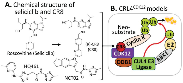
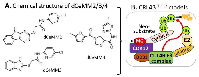
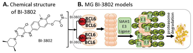
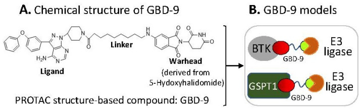
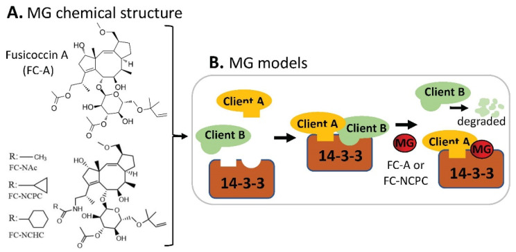
Molecular glue (MG) compounds are a type of unique small molecule that can change the protein-protein interactions (PPIs) and interactomes by degrading, stabilizing, or activating the target protein after their binging. These small-molecule MGs are gradually being recognized for their potential application in treating human diseases, including cancer. Evidence suggests that small-molecule MG compounds could essentially target any proteins, which play critical roles in human disease etiology, where many of these protein targets were previously considered undruggable. Intriguingly, most MG compounds with high efficacy for cancer treatment can glue on and control multiple key protein targets. On the other hand, a single key protein target can also be glued by multiple MG compounds with distinct chemical structures. The high flexibility of MG-protein interaction profiles provides rich soil for the growth and development of small-molecule MG compounds that can be used as molecular tools to assist in unraveling disease mechanisms, and they can also facilitate drug development for the treatment of human disease, especially human cancer. In this review, we elucidate this concept by using various types of small-molecule MG compounds and their corresponding protein targets that have been documented in the literature.

Keywords: E3 ligase protein complex; cancer; human disease; molecular glue (MG); neurodegenerative disease; protein degradation; protein ubiquitination; small-molecule compounds.


Conflict of interest statement

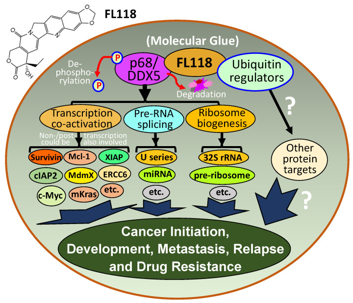
FL118 and FL118 core structure-based analogs will be further developed in Canget BioTekpharma LLC (www.canget-biotek.com), a spinoff company from Roswell Park Comprehensive Cancer Center (www.roswellpark.org). FL and XL are two of the eighteen initial investors of Canget for the development of FL118 and FL118 core structure platform-relevant anticancer agents.

Figures

Figure 7
Figure 7
(A) Chemical structure of the velcrin-type of MG compounds; (B) DNMDP-mediated PDE3A–SLFN12 heterotetramer formation models; (C) a cartoon model for elucidating the velcrin compound-induced mechanism of action for inducing cell death; (D) summary of the structure–activity relationship (SAR) of representative anagrelide analogs for deriving the lead compound A6. (D) is adapted from an original publication by Chen et al. [33].
Figure 1
Figure 1
(A) The IMiD-type MG compounds’ chemical structures; (B) the substrate receptor CRBN-involved E3 ligase protein complex model. Each compound in (A) was documented to engage the same E3 ligase protein complex (CRL4CRBN) to polyubiquitinate the compound’s neosubstrates shown in Table 1. Then, the polyubiquitinated neosubstrates/protein targets in Table 1 would be degraded through the ubiquitination proteasome pathway; and (C) chemical structure of FPJFT-2216-derived three new small molecules (TMX-4100, TMX-4113, TMX-4116).
Figure 2
Figure 2
This diagram shows the mode of action of folate–pomalidomide and folate-conjugated pomalidomide-based PROTACs. Upon binding FOLR1 on the cell membrane (1), folate-pomalidomide or folate-conjugated IMiD-based PROTACs are transported into cells, and the active pomalidomide or PROTACs are released after the reduction by endogenous GSH (2). The active pomalidomide or PROTACs recruit endogenous CRBN E3 ligase (similar to the model shown in Figure 1B), leading to polyubiquitination and subsequent degradation of the glued proteins of interest (POIs, neosubstrates) by the UPS (3). This figure is adapted from an original paper by Chen et al. [14].
Figure 3
Figure 3
(A) The aryl sulfonamide type MG compounds’ chemical structures; (B) the substrate receptor DCAF15-involved E3 ligase protein complex model. Each compound in (A) was documented to engage the same E3 ligase protein complex (CRL4DCAF15) to polyubiquitinate the compound’s neosubstrates shown in Table 2. Then, the polyubiquitinated neosubstrates/protein targets in Table 2 would be degraded through the ubiquitination proteasome pathway.
Figure 4
Figure 4
(A) The chemical structure of roscovitine/Seliciclib, CR8, HQ461, and NCT02 as MG compounds; (B) the substrate receptor-independent E3 ligase protein complex model for ubiquitination of the neosubstrate cyclin K. The MG compound CR8 shown in (A) was found through a substrate receptor-independent manner (neither CRBN nor DCAF15 being involved) to glue CDK12–cyclin K directly on DDB1–CUL4 E3 ligase complex to polyubiquitinate cyclin K. Then, the polyubiquitinated cyclin K would be degraded through the ubiquitination proteasome pathway. HQ461 and NCT02 may use a mechanism similar to CR8.
Figure 5
Figure 5
(A) The chemical structure of dCeMM2, dCeMM3, and dCeMM4 as MG compounds; (B) the substrate receptor-independent E3 ligase protein complex model for ubiquitination of the neosubstrate cyclin K. The MG compounds dCeMM2/3/4 shown in (A) was found through a substrate receptor-independent manner (neither CRBN nor DCAF15 being involved) to glue CDK12-cyclin K directly on DDB1–CUL4B E3 ligase complex to polyubiquitinate cyclin K. Then, the polyubiquitinated cyclin K would be degraded through the ubiquitination proteasome pathway.
Figure 6
Figure 6
(A) The chemical structure of BI-3802 as a special MG compound; (B) the diagram model for BI-3802-induced BCL6 polymerization, ubiquitination, and degradation. BI-3802 through the BTB domain of BCL6 makes BCL6 dimerization and then induces the dimerized BCL6 formation of helical filament. The polymerized BCL6 enhances the E3 ligase SIAHI interaction and ubiquitination of the polymerized BCL6 protein for degradation through the ubiquitination proteasome pathway.
Figure 8
Figure 8
(A) Chemical structure of the GBD-9 compound; (B) cartoon models for the GBD-9 ligand part to find two distinct neosubstrate proteins (BTK, GSPT1) for polyubiquitination by E3 ligases. Then, the polyubiquitinated proteins would be degraded through the ubiquitination proteasome pathway.
Figure 9
Figure 9
(A) Cartoon models of CDC34A and E3 ligase-mediated substrate ubiquitination being blocked by MG CC0651 (i.e., inhibition of ubiquitin transfer by stabilizing the noncovalent CDC34A-donor ubiquitin complex); (B) schematic of TR-FRET assay to detect CDC34A/UbE2R1–ubiquitin interactions. The assay used an N-terminal His-tagged CDC34A for recognition by an anti-His6 antibody coupled to Tb3+ and an N-terminal cysteine mutant of ubiquitin (denoted UbCys0) stoichiometrically labeled with 5′-iodoacetamide-fluorescein. In response to titration with CC0651, excitation of Tb3+ at 340 nm resulted in fluorescence energy transfer to the fluorescein moiety and emission at 520 nm; (C) diagram to explain the led compounds 2ab, 2cb, 2db, 2gb, and 2aη generated by medicinal chemistry hybridization of the prototype compound CC0651 and the isonipecotamide hit BDC22455743; (D) Ub IC50 (µM), TR-FRET EC50 (µM), and p27-Ub(n) IC50 (µM) for the prototype compound CC0651 and the led compounds 2ab, 2cb, 2db, 2gb, and 2aη were shown. IC50 values represent the mean +/− variance, n = 2. EC50 values represent the mean +/− SD, n = 3. (B,D) are adapted from an original paper by St-Cyr et al. (some information is also from their provided supplemental materials) [37].
Figure 10
Figure 10
(A) Chemical structure of the non-canonical MG compound asukamycin; (B) asukamycin-mediated covalently engaged TP53/p53-UBR7 formation models.
Figure 11
Figure 11
FL118 mechanism of action (MOA). DDX5 is a multifunctional master regulator involved in (1) co-activation of transcription of many oncogenes through the direct interactions of different transcription factors (e.g., c-Myc) in the oncogenic gene promoters, (2) regulation of miRNA and pre-RNA splicing (e.g., U1, U2, U3, …, snRNP), and (3) ribosome biogenesis (e.g., 32S rRNA, pre-ribosome). The small-molecule drug FL118 directly binds to and functionally dephosphorylates and degrades DDX5 protein (without decreasing DDX5 mRNA) through the proteasome degradation pathway. This suggests that FL118 could glue both DDX5 and ubiquitin-involved protein stability/degradation regulators (i.e., FL118 acts as a “molecular glue degrader”). All the DDX5 downstream protein targets were known to be involved in cancer initiation, development, metastasis, recurrence, and treatment resistance. Therefore, indirectly blocking DDX5 downstream targets through direct dephosphorylation and degradation of DDX5 by FL118 could result in FL118 high antitumor efficacy as demonstrated in our recent study, which used human CRC and PDAC cell and tumor models [49].
Figure 12
Figure 12
(A) Chemical structure of AN1, AN2, 10O5, and 8F20 as MG compounds; (B) a cartoon model to show the key finding for disease treatment. Such types of MG discovered by the logically designed assay could only ligate LC3 to mHTT but not wtHTT. Thus, mHTT could be glued on LC3 for degradation of mHTT but not wtHTT by autophagy.
Figure 13
Figure 13
(A) Chemical structure of fusicoccin A (FC-A), FC-NAc, FC-NCPC, and FC-NCHC as MG compounds; (B) a cartoon model to show the key finding for potential disease treatment. This type of MG compound could strengthen some 14-3-3 clients’ binding to 14-3-3, while disrupting other clients’ binding on 14-3-3 as shown in (B).

Similar articles

Cited by

References

    1. Faux M.C., Scott J.D. Molecular glue: Kinase anchoring and scaffold proteins. Cell. 1996;85:9–12. doi: 10.1016/S0092-8674(00)81075-2. - DOI - PubMed
    1. Schreiber S.L. The Rise of Molecular Glues. Cell. 2021;184:3–9. doi: 10.1016/j.cell.2020.12.020. - DOI - PubMed
    1. Tan X., Calderon-Villalobos L.I., Sharon M., Zheng C., Robinson C.V., Estelle M., Zheng N. Mechanism of auxin perception by the TIR1 ubiquitin ligase. Nature. 2007;446:640–645. doi: 10.1038/nature05731. - DOI - PubMed
    1. Sheard L.B., Tan X., Mao H., Withers J., Ben-Nissan G., Hinds T.R., Kobayashi Y., Hsu F.F., Sharon M., Browse J., et al. Jasmonate perception by inositol-phosphate-potentiated COI1-JAZ co-receptor. Nature. 2010;468:400–405. doi: 10.1038/nature09430. - DOI - PMC - PubMed
    1. Ito T., Ando H., Suzuki T., Ogura T., Hotta K., Imamura Y., Yamaguchi Y., Handa H. Identification of a primary target of thalidomide teratogenicity. Science. 2010;327:1345–1350. doi: 10.1126/science.1177319. - DOI - PubMed

MeSH terms