. 2022 Jun 2;23(11):6233.
doi: 10.3390/ijms23116233.

Signaling Pathways Used by the Specialized Pro-Resolving Mediator Maresin 2 Regulate Goblet Cell Function: Comparison with Maresin 1

Affiliations

Signaling Pathways Used by the Specialized Pro-Resolving Mediator Maresin 2 Regulate Goblet Cell Function: Comparison with Maresin 1

Markus V Olsen et al. Int J Mol Sci. .

Abstract

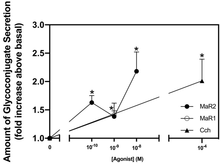
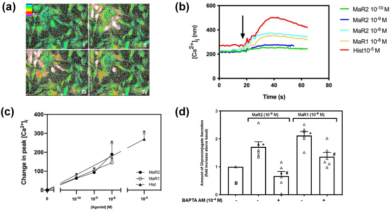
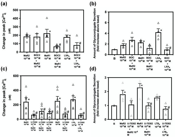
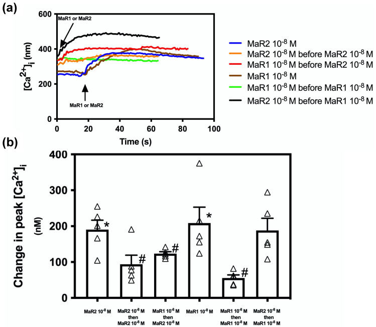
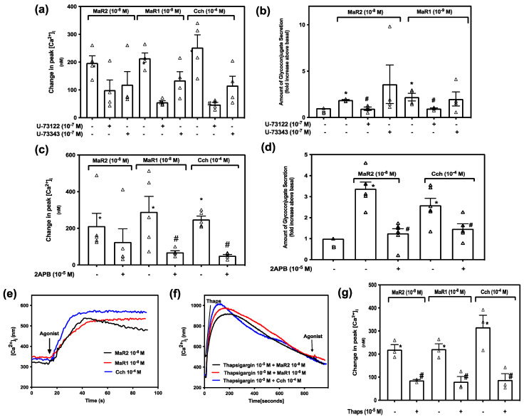
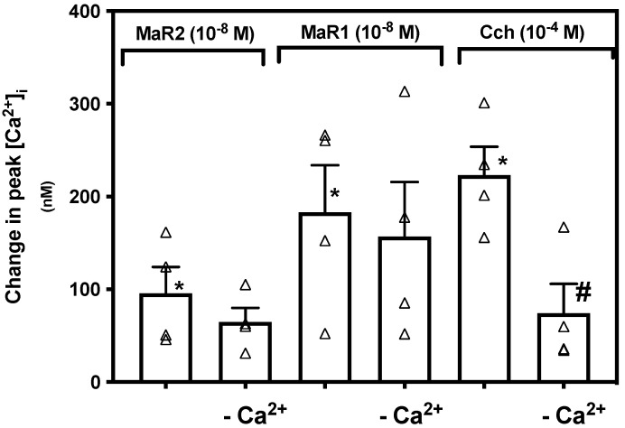
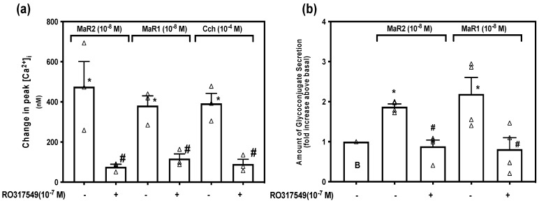
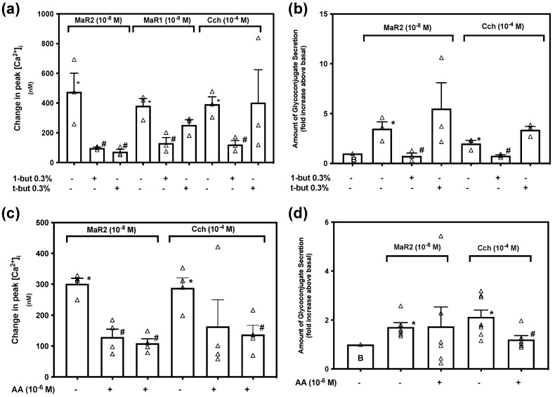
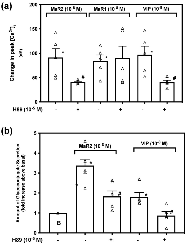
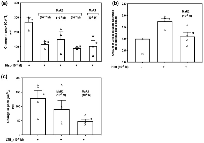
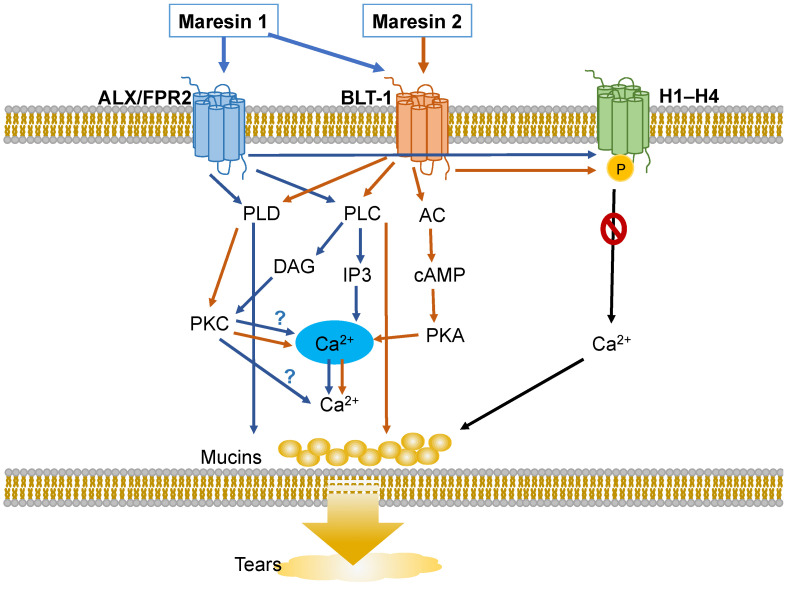
Specialized pro-resolving mediators (SPMs), including Maresins (MaR)-1 and 2, contribute to tear film homeostasis and resolve conjunctival inflammation. We investigated MaR2's signaling pathways in goblet cells (GC) from rat conjunctiva. Agonist-induced [Ca2+]i and high-molecular weight glycoconjugate secretion were measured. MaR2 increased [Ca2+]i and stimulated secretion. MaR2 and MaR1 stimulate conjunctival goblet cell function, especially secretion, by activating different but overlapping GPCR and signaling pathways, and furthermore counter-regulate histamine stimulated increase in [Ca2+]i. Thus, MaR2 and MaR1 play a role in maintaining the ocular surface and tear film homeostasis in health and disease. As MaR2 and MaR1 modulate conjunctival goblet cell function, they each may have potential as novel, but differing, options for the treatment of ocular surface inflammatory diseases including allergic conjunctivitis and dry eye disease. We conclude that in conjunctival GC MaR2 and MaR1, both increase the [Ca2+]i and stimulate secretion to maintain homeostasis by using one set of different, but overlapping, signaling pathways to increase [Ca2+]i and another set to stimulate secretion. MaR2 also resolves ocular allergy.

Keywords: epithelial cell; eye; inflammation; intracellular Ca2+; mucin secretion; tear film.


Conflict of interest statement

The authors declare no conflict of interest. The funders had no role in the design of the study; in the collection, analyses, or interpretation of data; in the writing of the manuscript, or in the decision to publish the results.

Figures

Figure 1
Figure 1
Maresin 2 (MaR2) stimulates glycoconjugate secretion. Rat conjunctival goblet cells were stimulated with either MaR2 (10−10–10−8 M) Maresin 1 (MaR1, 10−8 M), or carbachol (Cch, 10−4 M) for 2 hr. High molecular weight glycoprotein secretion was measured. Data are mean ± SEM from six experiments. * shows significance above basal. SEM, standard error of the mean.
Figure 2
Figure 2
Maresin 2 increases [Ca2+]i in rat conjunctival goblet cells. Pseudocolor images of rat conjunctival goblet cells stimulated with MaR2 (10−8 M) are shown in (a). MaR1 (10−8 M) is shown at four different times of stimulation. Panel I shows rat conjunctival goblet cell baseline [Ca2+]i level before stimulation (AI), panel II 30 s after stimulation with MaR2 (10−8 M) (AII); panel III 50 s after stimulation (AIII); and panel IV 80 s after stimulation (AIV). Changes in [Ca2+]i over time at different concentrations of MaR2 (10−8 M) are shown in (b). Changes in peak [Ca2+]i after stimulation with MaR2 (10−8 M) are shown in (c) Goblet cells were preincubated with BAPTA/AM (10−4 M) and stimulated with MaR2 (10−8 M) or MaR1 (10−9 M) for glycoconjugate secretion (d). Data are mean ± SEM from three (b,c) and six (d) experiments). White triangles indicate individual data points. * shows significance above zero (basal). # shows significant difference between agonist and agonist + inhibitor.
Figure 3
Figure 3
Inhibition of the BLT1, but not the ALX/FPR2 receptors acts on stimulation of [Ca2+]i and secretion by MaR2. Goblet cells were treated with the ALX/FPR2 receptor inhibitor BOC2 (10−4 M) for 30 min and stimulated with MaR2 10−8 M (a,b), MaR1 10−8 M (a) MaR1 10−9 M (b), LXA4 10−9 M (a) or LXA4 10−8 M (b). Goblet cells were treated with the BLT1 receptor inhibitor U-75302 (10−8–10−6 M) for 30 min and stimulated with MaR2 10−8 M (c,d), MaR1 10−8 M (c), MaR1 10−9 M (d) or LTB4 10−9 M (c) LTB4 10−8 M (d). [Ca2+]i was measured in (a,c); secretion in (b,d). Data are mean ± SEM of five (a), four (b), six (c) and four (d) experiments. White triangles indicate individual data points. * shows significance above basal. # shows significance between agonist and inhibitor + agonist.
Figure 4
Figure 4
Maresin 2 does not inhibit Maresin 1-stimulated increase in [Ca2+]i, but Maresin 1 does inhibit Maresin 2 stimulation. (a) shows changes in [Ca2+]i with time, while (b) shows changes in peak of [Ca2+]i. Goblet cells were preincubated 2 min with either MaR2 (10−8 M) (blue line a, first bar b) or MaR1 (10−8 M) (brown line a, fourth bar b), then stimulated with MaR2 (10−8 M) (orange line a, second bar b) or MaR1 (10−8 M (green line a, fifth bar b), respectively or stimulated with MaR1 before MaR2 (red line a, third bar b) or MaR2 before MaR1 (black line a, (sixth bar b). Data are mean ± SEM of five (a and b) experiments. White triangles indicate individual data points. * shows significance above basal. # shows significance between MaR2 and MaR2 after MaR2 or MaR1, and between MaR1 and MaR1 after MaR2 or MaR1.
Figure 5
Figure 5
Maresin 2 increase in [Ca2+]i, but not secretion, is independent of the PLC-pathway in rat conjunctival goblet cells. Goblet cells were treated with vehicle, the active PLC inhibitor U-73122 or the inactive PLC inhibitor U-73343 both at 10−7 M for 30 min and stimulated with MaR2 (10−8 M), MaR1 (10−8 M) or Cch (10−4 M). (a) shows changes in peak [Ca2+]i and (b) shows glycoconjugate secretion. Goblet cells were treated with vehicle or 2APB (10−5 M) and stimulated with MaR2 (10−8 M), MaR1 (10−8 M) or Cch (10−4 M). (c) shows changes in peak [Ca2+]i, and (d) shows glycoconjugate secretion. Goblet cells were stimulated with MaR2 (10−8 M), MaR1 (10−8 M) or Cch (10−4 M) alone or incubated with vehicle or thapsigargin (10−5 M) for 15 min and then stimulated with MaR2 (10−8 M), MaR1 (10−8 M) or Cch (10−4 M). (e,f) show changes in [Ca2+]i with time, while (g) shows changes in peak of [Ca2+]i. Data are mean ± SEM of four (a), four (b), five (c), six (d), and three (eg) experiments. White triangles indicate individual data points. * shows significance above basal. # shows significance between MaR2 and inhibitor then MaR2 or control and inhibitor then control.
Figure 6
Figure 6
Maresin 2 stimulated increase in [Ca2+]i is independent of extracellular Ca2+ in rat conjunctival goblet cells. Goblet cells were incubated with vehicle with or without CaCl2, then stimulated with MaR2 (10−8 M), or the controls MaR1 (10−8 M) and Cch (10−4 M). Figure shows changes in peak of [Ca2+]i. Data are mean ± SEM of four experiments. White triangles indicate individual data points. * shows significance above basal. # shows significance between agonist and extracellular Ca2+ removal then agonist.
Figure 7
Figure 7
Maresin 2 increases [Ca2+]i and stimulates secretion by activation of protein kinase C (PKC). Goblet cells were incubated with RO317549 (10−7 M) for 30 min, then stimulated with MaR2 (10−8 M) or the positive controls MaR1 (10−8 M) or Cch (10−4 M) and then [Ca2+]i and secretion were measured. (a) shows change in peak [Ca2+]i, while (b) shows -fold increase in glycoconjugate secretion. Data are mean ± SEM of three (a) and four (b) experiments. White triangles indicate individual data points. * shows significance above basal. # shows significance between agonist and inhibitor then agonist.
Figure 8
Figure 8
Maresin 2 has different dependency on Phospholipase D (PLD) compared to Phospholipase A2 (PLA2) to increase [Ca2+]i and stimulate secretion in rat conjunctival goblet cells. Goblet cells were preincubated with the PLD inhibitor 0.3% 1-butanol or the inactive analog 0.3% t-butanol for 15 min and then stimulated with MaR2 (10−8 M), MaR1 (10−8 M) or Cch (10−4 M) to measure the change in peak [Ca2+]i (a), or secretion (b). Goblet cells were preincubated with the PLA2 inhibitor aristolochic acid 10−5 M or 10−6 M for 30 min and stimulated with MaR2 (10−8 M) or Cch (10−4 M) to measure the change in peak [Ca2+]i (c), or secretion (d). Data are mean ± SEM of three (a), three (b), four (c) and six (d) experiments. White triangles indicate individual data points. * shows significance above basal. # shows significance between agonist and inhibitor followed by agonist.
Figure 9
Figure 9
Maresin 2 uses protein kinase A to increase [Ca2+]i and stimulate secretion in rat conjunctival goblet cells. Goblet cells were incubated with the protein kinase A (PKA) inhibitor H89 (10−5 M) for 30 min and then stimulated with MaR2 (10−8 M) or the positive controls MaR1 (10−8 M) and VIP (10−8 M) to measure the change in peak [Ca2+]i (a), or secretion (b). Note that MaR1 was not used as an agonist in (b). Data are mean ± SEM of five (a) and six (b) experiments. White triangles indicate individual data points. * shows significance above basal. # shows significance between agonist and inhibitor followed by agonist.
Figure 10
Figure 10
Maresin 2 inhibits histamine-, but not LTB4-stimulated increase in [Ca2+]i and histamine-stimulated glycoconjugate secretion in rat conjunctival goblet cells. Goblet cells were incubated with MaR2 (10−8 M) and then stimulated with histamine (10−5 M). Goblet cells were incubated with MaR2 (10−10 to 10−8 M) or MaR1 (10−8 M) for 30 min, then stimulated with histamine (10−5 M). Changes in in peak [Ca2+]i are shown in (a) and changes in glycoconjugate secretion are shown in (b). Goblet cells were incubated with MaR2 (10−8 M) or MaR1 (10−8 M) for 30 min, then stimulated with LTB4 10−9 M. Changes in peak [Ca2+]i are shown in (c). Data are mean ± SEM of three (a) and four (b) and four (c) experiments. White triangles indicate individual data points. * shows significance above basal. # shows significance between MaR2 or MaR1 treatment and histamine (a,b) or LTB4 (c) and histamine (a,b) or LTB4 (c) alone.
Figure 11
Figure 11
Schematic diagram of signaling pathways activated by Maresin 2 (MaR2) (orange arrows) compared to the pathways activated by Maresin 1 (MaR1) (blue arrows). MaR2 activates the BLT1 receptor activating PLD, AC, PLA2, and PLC. PLD and AC activate downstream molecules that increase [Ca2+]i causing glycoprotein secretion. PLA2 and the PLC pathway stimulates glycoprotein secretion by another unknown mechanism than increasing [Ca2+]i.

Similar articles

Cited by

References

    1. Hodges R.R., Dartt D.A. Tear film mucins: Front line defenders of the ocular surface; comparison with airway and gastrointestinal tract mucins. Exp. Eye Res. 2013;117:62–78. doi: 10.1016/j.exer.2013.07.027. - DOI - PMC - PubMed
    1. Dartt D.A. Control of mucin production by ocular surface epithelial cells. Exp. Eye Res. 2004;78:173–185. doi: 10.1016/j.exer.2003.10.005. - DOI - PubMed
    1. Argueso P., Balaram M., Spurr-Michaud S., Keutmann H.T., Dana M.R., Gipson I.K. Decreased levels of the goblet cell mucin MUC5AC in tears of patients with Sjogren syndrome. Investig. Ophthalmol. Vis. Sci. 2002;43:1004–1011. - PubMed
    1. Dogru M., Matsumoto Y., Okada N., Igarashi A., Fukagawa K., Shimazaki J., Tsubota K., Fujishima H. Alterations of the ocular surface epithelial MUC16 and goblet cell MUC5AC in patients with atopic keratoconjunctivitis. Allergy. 2008;63:1324–1334. doi: 10.1111/j.1398-9995.2008.01781.x. - DOI - PubMed
    1. Zhao H., Jumblatt J.E., Wood T.O., Jumblatt M.M. Quantification of MUC5AC protein in human tears. Cornea. 2001;20:873–877. doi: 10.1097/00003226-200111000-00019. - DOI - PubMed

Substances