. 2022 Jun 2;23(11):6260.
doi: 10.3390/ijms23116260.

Venetoclax Induces Cardiotoxicity through Modulation of Oxidative-Stress-Mediated Cardiac Inflammation and Apoptosis via NF-κB and BCL-2 Pathway

Affiliations

Venetoclax Induces Cardiotoxicity through Modulation of Oxidative-Stress-Mediated Cardiac Inflammation and Apoptosis via NF-κB and BCL-2 Pathway

Abdullah F AlAsmari et al. Int J Mol Sci. .

Abstract

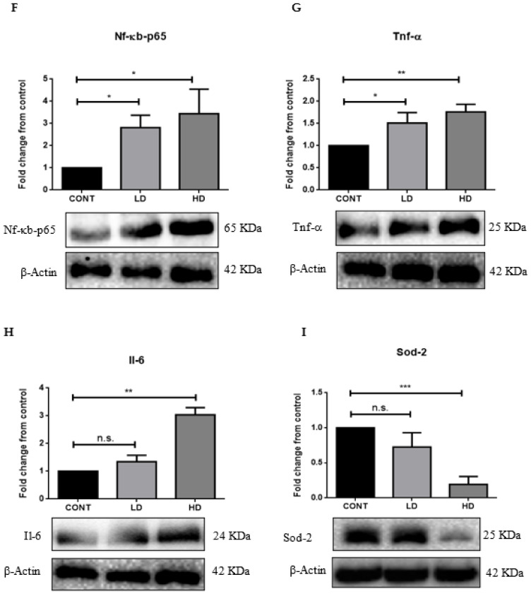
Cardiovascular damage induced by anticancer therapy has become the main health problem after tumor elimination. Venetoclax (VTX) is a promising novel agent that has been proven to have a high efficacy in multiple hematological diseases, especially acute myeloid leukemia (AML) and chronic lymphocytic leukemia (CLL). Considering its mechanism of action, the possibility that VTX may cause cardiotoxicity cannot be ruled out. Therefore, this study was designed to investigate the toxic effect of VTX on the heart. Male Sprague-Dawley rats were randomly divided into three groups: control, low-dose VTX (50 mg/kg via oral gavage), and high-dose VTX (100 mg/kg via oral gavage). After 21 days, blood and tissue samples were collected for histopathological, biochemical, gene, and protein analyses. We demonstrated that VTX treatment resulted in cardiac damages as evidenced by major changes in histopathology and markedly elevated cardiac enzymes and hypertrophic genes markers. Moreover, we observed a drastic increase in oxidative stress, as well as inflammatory and apoptotic markers, with a remarkable decline in the levels of Bcl-2. To the best of our knowledge, this study is the first to report the cardiotoxic effect of VTX. Further experiments and future studies are strongly needed to comprehensively understand the cardiotoxic effect of VTX.

Keywords: apoptosis; cardiotoxicity; inflammation; oxidative stress; venetoclax.


Conflict of interest statement

The authors declare no conflict of interest.

Figures

Figure 1
Figure 1
Serum levels of cardiac enzymes. Sera from different groups were analyzed to measure CK-MB and cTnI levels (A,B, respectively). Data are presented as mean ± SD (n = 5). * p < 0.05. Cont, control; LDV, low-dose venetoclax (50 mg/kg); HDV, high-dose venetoclax (100 mg/kg); CK-MB, creatine kinase-MB; cTn-I, troponin I cardiac muscle.
Figure 2
Figure 2
VTX-induced cardiac hypertrophy. (A) Body weight; (B) heart weight; and (C) heart weight to body weight ratio. Data are presented as mean ± SD. * p < 0.05; n.s., no significant changes were observed (p > 0.05). (DG) mRNA levels of (D) α-Mhc, (E) β-Mhc, (F) β-Mhc to α-Mhc ratio, and (G) Bnp were measured using RT-PCR. Data are presented as mean ± SD (n = 5). * p < 0.05, ** p < 0.01; n.s., no significant changes were observed (p > 0.05). Data were normalized to β-actin as a housekeeping gene and one-way analysis of variance (ANOVA), followed by Tukey–Kramer multiple-comparisons tests. Cont, control; LD, low-dose venetoclax (50 mg/kg); HD, high-dose venetoclax (100 mg/kg); α-Mhc, alpha myosin heavy chain; α-Mhc, beta myosin heavy chain; Bnp, brain natriuretic peptide.
Figure 3
Figure 3
Light micrographs of cardiac tissues using H&E stain. (A,D) Represents the regular, parallel, and branching striated myocardial fibers with cross-striation and regular nuclei with no evidence of inflammation or necrosis. (B,E) Myocardium section obtained from low-dose VTX shows normal appearance of the myocardial fibers, but with slightly nuclear enlargement. (C,F) Represents heart section from rats treated with a high dose of VTX, and shows the presence of a focus of myocardial damage associated with chronic inflammatory reaction (arrowhead). Images (AC) obtained at 400× magnification, while images (DF) were obtained at 600× magnification (scale bar = 50 µm and 20 µm, respectively). (G) Histopathological score. (H) Cardiomyocytes’ cross-sectional area. Data are presented as mean ± SD (n = 3). * p < 0.05, ** p < 0.01, *** p < 0.001. Cont, control; LD, low-dose venetoclax (50 mg/kg); HD, high-dose venetoclax (100 mg/kg).
Figure 4
Figure 4
VTX-induced apoptosis in the heart. The mRNA levels of Bax (A) and Bcl-2 (B) were measured using RT-PCR. Data were normalized to β-actin as a housekeeping gene and one-way analysis of variance (ANOVA), followed by Tukey–Kramer multiple-comparisons tests. (C) Representative Western blot analysis of Bcl-2 protein levels. (D) Representative Western blot analysis of Cleaved Cas-3 protein levels. Data are presented as mean ± SD (n = 5). * p < 0.05, ** p < 0.01; n.s., no significant changes were observed (p > 0.05). Cont, control; LD, low-dose venetoclax (50 mg/kg); HD, high-dose venetoclax (100 mg/kg); Bax, Bcl-2 associated X; Bcl-2, B-cell lymphoma 2; Cleaved Cas-3, cystinyl aspartate-specific proteases 3; β-actin, beta actin.
Figure 5
Figure 5
VTX-induced oxidative stress and inflammation in the heart. (AE) The mRNA levels of Ifn-γ, Tgf-β, Nf-κb-p-65, Tnf-α, and Il-6 were measured using RT-PCR. Data were normalized to β-actin as a housekeeping gene and one-way analysis of variance (ANOVA), followed by Tukey–Kramer multiple-comparisons tests. (FI) Representative Western blot analysis of protein levels of Nf-κb-p-65, Tnf-α, Il-6, and Sod-2. Data are presented as mean ± SD (n = 5). * p < 0.05, ** p < 0.01, *** p < 0.001; n.s., no significant changes were observed (p > 0.05). Cont, control; LD, low-dose venetoclax (50 mg/kg); HD, high-dose venetoclax (100 mg/kg); Ifn-γ, interferon gamma; Tgf-β, transforming growth factor beta; Nf-κb-p-65, nuclear factor kappa-B; Tnf-α, tumor necrosis factor alpha; Il-6, interleukin-6; Sod-2, superoxide dismutase-2; β-actin, beta actin.
Figure 5
Figure 5
VTX-induced oxidative stress and inflammation in the heart. (AE) The mRNA levels of Ifn-γ, Tgf-β, Nf-κb-p-65, Tnf-α, and Il-6 were measured using RT-PCR. Data were normalized to β-actin as a housekeeping gene and one-way analysis of variance (ANOVA), followed by Tukey–Kramer multiple-comparisons tests. (FI) Representative Western blot analysis of protein levels of Nf-κb-p-65, Tnf-α, Il-6, and Sod-2. Data are presented as mean ± SD (n = 5). * p < 0.05, ** p < 0.01, *** p < 0.001; n.s., no significant changes were observed (p > 0.05). Cont, control; LD, low-dose venetoclax (50 mg/kg); HD, high-dose venetoclax (100 mg/kg); Ifn-γ, interferon gamma; Tgf-β, transforming growth factor beta; Nf-κb-p-65, nuclear factor kappa-B; Tnf-α, tumor necrosis factor alpha; Il-6, interleukin-6; Sod-2, superoxide dismutase-2; β-actin, beta actin.
Figure 6
Figure 6
Effects of VTX on oxidative stress status. Biochemical analysis of MDA (A), CAT (B), and GSH (C). Data are presented as mean ± SD (n = 5). * p < 0.05, ** p < 0.01, *** p < 0.001; n.s., no significant changes were observed (p > 0.05). Cont, control; LD, low-dose venetoclax (50 mg/kg); HD, high-dose venetoclax (100 mg/kg); MDA, malondialdehyde; CAT, catalase; GSH, glutathione.
Figure 7
Figure 7
Schematic representation of cardiotoxicity mechanism of venetoclax (VTX).

References

    1. Laverty H.G., Benson C., Cartwright E.J., Cross M.J., Garland C., Hammond T., Holloway C., McMahon N., Milligan J., Park B.K., et al. How can we improve our understanding of cardiovascular safety liabilities to develop safer medicines? Br. J. Pharmacol. 2011;163:675–693. doi: 10.1111/j.1476-5381.2011.01255.x. - DOI - PMC - PubMed
    1. Cardinale D.M., Zaninotto M., Cipolla C.M., Passino C., Plebani M., Clerico A. Cardiotoxic effects and myocardial injury: The search for a more precise definition of drug cardiotoxicity. Clin. Chem. Lab. Med. 2020;59:51–57. doi: 10.1515/cclm-2020-0566. - DOI - PubMed
    1. Shah C.P., Moreb J.S. Cardiotoxicity due to targeted anticancer agents: A growing challenge. Ther. Adv. Cardiovasc. Dis. 2019;13:1753944719843435. doi: 10.1177/1753944719843435. - DOI - PMC - PubMed
    1. Kim H., Chung W.B., Im Cho K., Kim B.J., Seo J.S., Park S.M., Kim H.J., Lee J.H., Kim E.K., Youn H.J. Diagnosis, Treatment, and Prevention of Cardiovascular Toxicity Related to Anti-Cancer Treatment in Clinical Practice: An Opinion Paper from the Working Group on Cardio-Oncology of the Korean Society of Echocardiography. J. Cardiovasc. Ultrasound. 2018;26:1–25. doi: 10.4250/jcu.2018.26.1.1. - DOI - PMC - PubMed
    1. Dong J., Chen H. Cardiotoxicity of Anticancer Therapeutics. Front. Cardiovasc. Med. 2018;5:9. doi: 10.3389/fcvm.2018.00009. - DOI - PMC - PubMed

LinkOut - more resources