. 2022 Jun 6;23(11):6351.
doi: 10.3390/ijms23116351.

Hypothalamic NPY-Y1R Interacts with Gonadal Hormones in Protecting Female Mice against Obesity and Neuroinflammation

Affiliations

Hypothalamic NPY-Y1R Interacts with Gonadal Hormones in Protecting Female Mice against Obesity and Neuroinflammation

Alessandra Oberto et al. Int J Mol Sci. .

Abstract

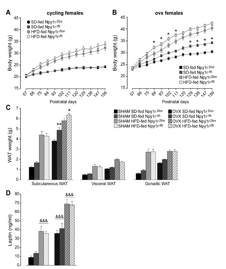
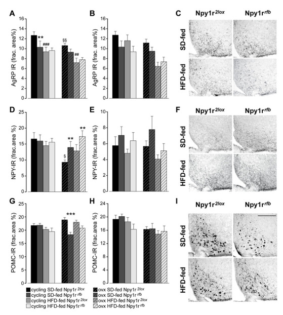
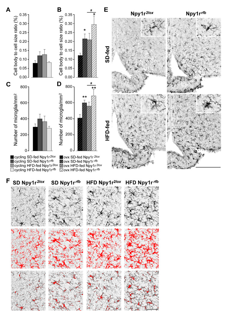
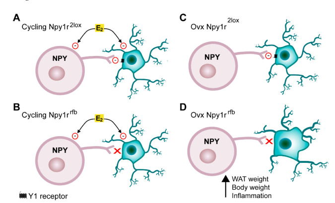
We previously demonstrated that Npy1rrfb mice, which carry the conditional inactivation of the Npy1r gene in forebrain principal neurons, display a sexually dimorphic phenotype, with male mice showing metabolic, hormonal and behavioral effects and females being only marginally affected. Moreover, exposure of Npy1rrfb male mice to a high-fat diet (HFD) increased body weight growth, adipose tissue, blood glucose levels and caloric intake compared to Npy1r2lox male controls. We used conditional knockout Npy1rrfb and Npy1r2lox control mice to examine whether forebrain disruption of the Npy1r gene affects susceptibility to obesity and associated disorders of cycling and ovariectomized (ovx) female mice in a standard diet (SD) regimen or exposed to an HFD for 3 months. The conditional deletion of the Npy1r gene increased body weight and subcutaneous white adipose tissue weight in both SD- and HFD-fed ovx females but not in cycling females. Moreover, compared with ovx control females on the same diet regimen, Npy1rrfb females displayed increased microglia number and activation, increased expression of Neuropeptide Y (NPY)-immunoreactivity (IR) and decreased expression of proopiomelanocortin-IR in the hypothalamic arcuate nucleus (ARC). These results suggest that in the ARC NPY-Y1R reduces the susceptibility to obesity of female mice with low levels of gonadal hormones and that this effect may be mediated via NPY-Y1R ability to protect the brain against neuroinflammation.

Keywords: high-fat diet; hypothalamic Y1R receptors; neuroinflammation; obesity; ovariectomy.


Conflict of interest statement

The authors declare no conflict of interest.

Figures

Figure 1
Figure 1
Body weight of standard diet (SD)- and high-fat diet (HFD)-fed cycling (A) and ovx (B) Npy1r2lox control and Npy1rrfb knockout female mice. No significant differences in body weight growth were observed between cycling Npy1r2lox and Npy1rrfb female mice during postnatal days (P) 57–156, independently of diet regimen. SD- and HFD-fed ovx Npy1rrfb females displayed a significantly greater body weight compared with their control littermates on the same diet regimen. Data are the mean ± SEM; n = 17–26 from 6 litters. * p < 0.05 versus the corresponding control females. (C) Adiposity of standard diet (SD)- and high-fat diet (HFD)-fed cycling and ovx Npy1r2lox control and Npy1rrfb knockout female mice. Significantly larger depots of subcutaneous fat were observed in ovx Npy1rrfb females compared with their control littermates on the same diet regimen. Data are the mean ± SEM; n = 10–15 from 3 litters. ** p < 0.01 and * p < 0.05 versus SD-fed and HFD-fed ovx Npy1r2lox female mice, respectively. (D) Leptin plasma levels of standard diet (SD)- and high-fat diet (HFD)-fed cycling and ovx Npy1r2lox control and Npy1rrfb knockout female mice. Both ovariectomy and HFD increased leptin plasma levels of female mice. Data are the mean ± SEM; n = 10–15 from 3 litters. &&& p < 0.001.
Figure 2
Figure 2
(A) Spontaneous locomotor activity of standard diet (SD)- and high-fat diet (HFD)-fed cycling and ovx Npy1r2lox control and Npy1rrfb knockout female mice. Both HFD and ovariectomy significantly decreased locomotor activity (distance traveled, cm) of Npy1r2lox and Npy1rrfb female mice. HFD-fed cycling and SD-fed ovx Npy1rrfb females showed significantly lower locomotor activity compared with their corresponding controls. Data are the mean ± SEM; n = 8–12 from 2 litters. # p < 0.05 versus SD-fed cycling controls; ### p < 0.001 versus SD-fed cycling Npy1rrfb female mice; § p < 0.05 versus SD-fed cycling controls; §§§ versus SD-fed cycling Npy1rrfb female mice; * p < 0.05 versus SD-fed ovx controls. (B) Correlation between locomotor activity and body weight of 156 days old SD- and HFD-fed cycling and ovx Npy1r2lox and Npy1rrfb female mice. Data are the mean ± SEM; n = 6 from 3 litters. Food and energy intake of standard diet (SD)- and high-fat diet (HFD)-fed cycling and ovx Npy1r2lox control and Npy1rrfb knockout female mice (CE). (C) No significant differences of cumulative food intake (g) were observed among the different experimental groups. Data are the mean ± SEM; n = 4 from 3 litters. (D) HFD increased cumulative energy intake (Kcal) of cycling and ovx female mice, independently of the genotype. Data are the mean ± SEM; n = 6 from 3 litters. &&& p < 0.001 versus SD-females. (E) Nine-day cumulative energy intake (kcal) of SD- and HFD-fed cycling and ovx Npy1r2lox control and Npy1rrfb knockout female mice during the metabolic challenge procedure (P66–156). Data are the mean ± SEM; n = 6 from 3 litters.
Figure 3
Figure 3
Glucose homeostasis and metabolism of standard diet (SD)- and high-fat diet (HFD)-fed cycling and ovx Npy1r2lox control and Npy1rrfb knockout female mice (AF). (A) Glucose tolerance tests and (B) area under the curve (AUC) of SD- and HFD-fed cycling and ovx Npy1r2lox and Npy1rrfb females. Data are the mean ± SEM; n = 11–20 from 3 litters. ## p < 0.01 HFD-fed versus SD-fed cycling controls; ### p < 0.001 HFD-fed versus SD-fed cycling Npy1r2lox female mice. (C) Insulin tolerance test and (D) area under the curve (AUC) of SD- and HFD-fed cycling and ovx Npy1r2lox and Npy1rrfb females. Data are the mean ± SEM; n = 7–15 from 2 litters. (E) Pyruvate tolerance test and (F) area under the curve (AUC) of SD- and HFD-fed cycling and ovx Npy1r2lox and Npy1rrfb females. Data are the mean ± SEM; n = 7–16 from 2 litters. # p < 0.05 HFD-fed versus SD-fed cycling Npy1rrfb females. (G) Heart rate and (H) blood pressure of standard diet (SD)- and high-fat diet (HFD)-fed cycling and ovx Npy1r2lox control and Npy1rrfb knockout female mice. &&& p < 0.001 versus cycling females.
Figure 4
Figure 4
Expression of neuropeptides in the ARC and PVN of standard diet (SD)- and high-fat diet (HFD)-fed cycling and ovx Npy1r2lox control and Npy1rrfb knockout female mice. Agouti-related protein immunoreactivity (AgRP-IR) in the ARC (A) and in the PVN (B) of SD- and HFD-fed cycling and ovx Npy1r2lox and Npy1rrfb females. HFD and ovx significantly decreased AgRP expression in the ARC of female mice. SD-fed cycling Npy1rrfb conditional mutants showed a significant decrease of AgRP-IR in the ARC compared to SD-fed cycling control females. Data are the mean ± SEM; n = 8–9 (ARC) and n = 7–10 (PVN) from 2 litters. ** p < 0.01 and ### p < 0.001 versus SD-fed cycling Npy1r2lox females; §§ p < 0.01 versus SD-fed cycling Npy1r2lox; mice; ## p < 0.01 versus SD-fed ovx Npy1r2lox female mice. (C) Representative images of the ARC of ovx females expressing AgRP-IR immunoreactivity (scale bar: 150 μm). Neuropeptide Y immunoreactivity (NPY-IR) in the ARC (D) and in the PVN (E) of SD- and HFD-fed cycling and ovx Npy1r2lox and Npy1rrfb females. A significant decrease of NPY-IR was observed in the ARC of ovx Npy1r2lox mice compared with cycling Npy1r2lox mice. A significant increase of NPY-IR was observed in the ARC of ovariectomized Npy1rrfb mice compared with their control littermates on the same diet regimen. § p < 0.05 versus cycling Npy1r2lox females on the same diet regimen. ** p < 0.01 versus ovx Npy1r2lox on the same diet regimen. Data are the mean ± SEM; n = 5–8 from 2 litters. (F) Representative images of the ARC of ovx females expressing NPY-IR immunoreactivity (scale bar: 150 μm). Proopiomelanocortin immunoreactivity (POMC-IR) in the ARC (G) and in the PVN (H) of SD- and HFD-fed cycling and ovx Npy1r2lox and Npy1rrfb females. A significant decrease of POMC-IR was observed in the ARC of SD-fed ovx Npy1rrfb mice compared with SD-fed ovx Npy1r2lox mice. Data are the mean ± SEM; n = 7–11 from 2 litters. *** p < 0.001 versus SD-fed ovx Npy1r2lox mice. (I) Representative images of the ARC of ovx females expressing POMC-IR (scale bar: 150 μm).
Figure 5
Figure 5
Effect of HFD and Npy1r gene deletion on microglial activation and microglia number. The cell body to cell size ratio (cb/c) of microglia (A,B) and the number of microglia/mm2 (C,D). HFD significantly increased cb/c (B) and microglia number (D) in the ARC of ovx females. Conditional inactivation of Npy1r gene increased cb/c (B) and microglia number (D) in the ARC of SD- and HFD-fed ovx mice. Data are the mean ± SEM; n = 4–6 from 2 litters. # p < 0.05 versus SD-fed ovx females. * p < 0.05 and ** p < 0.01 versus Npy1r2lox ovx females on the same diet regimen. (E) Representative images of the ARC of ovx females expressing ionized calcium-binding adaptor protein-1 (IBA-1) immunoreactivity (scale bar: 150 μm). (F) Image analysis used to quantify the morphological characteristics of microglia in Iba-1 staining in ovx females. Upper panels: the unprocessed pictures. Middle panels: pixels darker than the background are traced (red) to determine the total cell size of microglia. Lower panels: pixel-clusters that are above an applied staining threshold and size-filter are traced (red) to determine the total cell body size of all microglia, as well as the number of microglia.
Figure 6
Figure 6
Interactions between estradiol and the NPY-Y1 receptor system in the ARC. (A) Estrogens (E2) directly inhibit NPY neurons. Microglia activation and proliferation is increased by HFD and inhibited by E2 and the NPY-Y1 receptor system. (B) In cycling females, the HFD-induced microglia activation is inhibited by E2 also in the absence of Y1 receptors on microglia. (C) In ovx Npy1r2lox females, NPY inhibits microglia through activation of Y1 receptors. (D) In Npy1rrfb ovx females, on both SD and HFD regimen, the deletion of Y1 receptors in microglia increases microglia activation and, in turn, body and WAT weight.

References

    1. Santos Machado V.d.S., Ribeiro Valadares A.L., Costa-Paiva L.H., Osis M.J., Sousa M.H., Pinto-Neto A.M. Aging, obesity, and multimorbidity in women 50 years or older: A population-based study. Menopause. 2013;20:818. doi: 10.1097/GME.0b013e31827fdd8c. - DOI - PubMed
    1. Wildman R.P., Tepper P.G., Crawford S., Finchelstein J.S., Sutton-Tyrrell K., Thurston R.C., Santoro N., Sternfeld B., Greendale G.A. Do changes in sex steroid hormones precede or follow increases in body weight during the menopause transition? Results from the Study of Women’s Health Across the Nation. J. Clin. Endocrinol. Metab. 2012;97:E1695–E704. doi: 10.1210/jc.2012-1614. - DOI - PMC - PubMed
    1. Lizcano F., Guzmán G. Estrogen deficiency and the origin of obesity during menopause. BioMed Res. Int. 2014;2014:757461. doi: 10.1155/2014/757461. - DOI - PMC - PubMed
    1. Della Torre S., Benedusi V., Fontana R., Maggi A. Energy metabolism and fertility—A balance preserved for female health. Nat. Rev. Endocrinol. 2014;10:13–23. doi: 10.1038/nrendo.2013.203. - DOI - PubMed
    1. Dorfman M.D., Krull J.E., Douglass J.D., Fasnacht R., Lara-Lince F., Meek T.H., Shi X., Damian V., Nguyen H.T., Matsen M.E., et al. Sex differences in microglial CX3CR1 signalling determine obesity susceptibility in mice. Nat. Commun. 2017;8:14556. doi: 10.1038/ncomms14556. - DOI - PMC - PubMed