

Pre-Interventional 3D-Printing-Assisted Planning of Flow Disrupter Implantation for the Treatment of an Intracranial Aneurysm
- PMID: 35683339
- PMCID: PMC9181068
- DOI: 10.3390/jcm11112950
Pre-Interventional 3D-Printing-Assisted Planning of Flow Disrupter Implantation for the Treatment of an Intracranial Aneurysm
Abstract
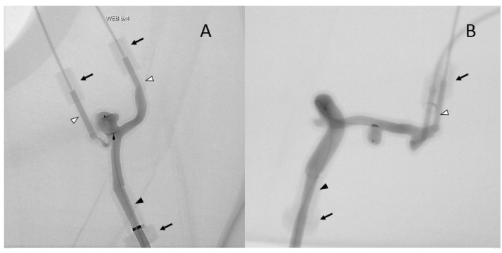
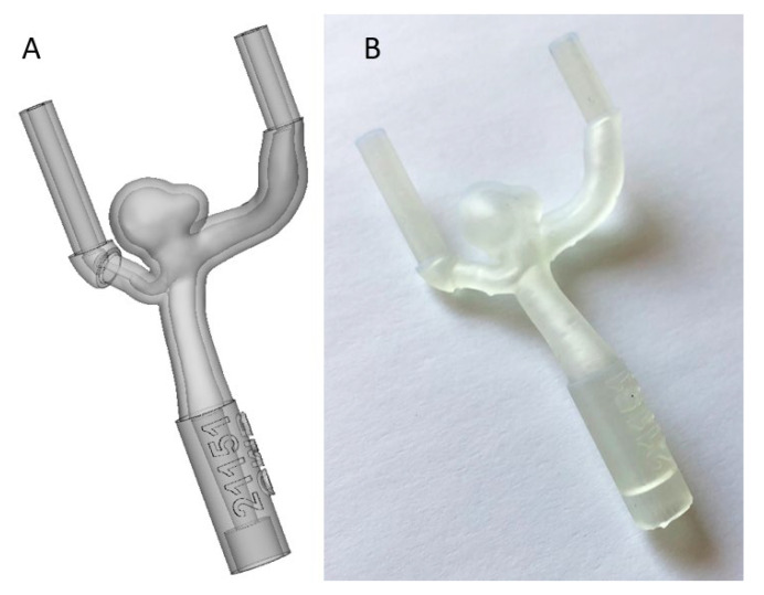
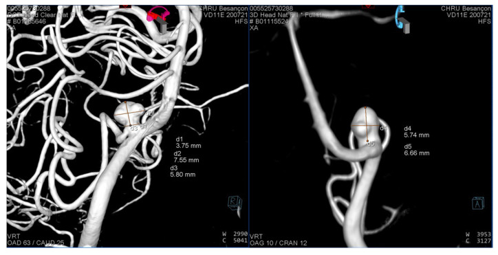
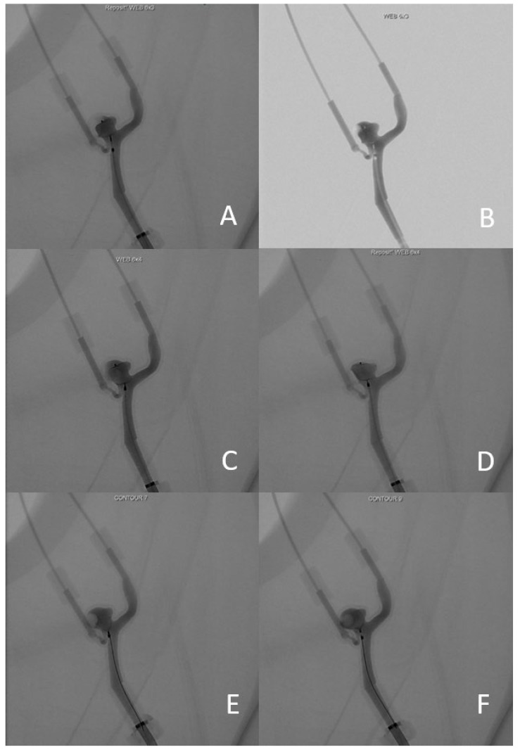
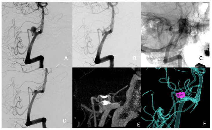
Intrasaccular flow disrupter devices (ISFD) have opened up new ways to treat intracranial aneurysms but choosing the correct size of ISFD can be challenging. We describe the first use of 3D printing to assist in the choice of ISFD, and we report an illustrative case. We developed a technique that uses preoperative angiography to make a plastic model of the aneurysm. We tested the deployment of different sizes of intrasaccular flow disruptor on the 3D model under fluoroscopy. The best devices were then used as the first-line strategy to treat the patient. The preoperative 3D printing helped in the successful selection of a first-line ISFD, which was not the one recommended by the manufacturer. Three-dimensional printing can provide interesting information regarding the treatment of intracranial aneurysms using ISFD. Further studies are needed to fully assess its benefits.
Keywords: 3D printing; aneurysm; contour; interventional neuroradiology; stroke; web.
Conflict of interest statement
The authors declare no conflict of interest. The funders had no role in the design of the study; in the collection, analyses, or interpretation of data; in the writing of the manuscript, or in the decision to publish the results.
Figures
References
-
- Piotin M., Biondi A., Sourour N., Mounayer C., Jaworski M., Mangiafico S., Andersson T., Söderman M., Goffette P., Anxionnat R., et al. The LUNA Aneurysm Embolization System for Intracranial Aneurysm Treatment: Short-Term, Mid-Term and Long-Term Clinical and Angiographic Results. J. Neurointerv. Surg. 2018;10:e34. doi: 10.1136/neurintsurg-2018-013767. - DOI - PMC - PubMed
-
- Cagnazzo F., Marnat G., Ferreira I., Daube P., Derraz I., Dargazanli C., Lefevre P.-H., Gascou G., Riquelme C., Morganti R., et al. Comparison of Woven EndoBridge Device Sizing with Conventional Measurements and Virtual Simulation Using the Sim&Size Software: A Multicenter Experience. J. NeuroInterv. Surg. 2021;13:924–929. doi: 10.1136/neurintsurg-2020-017060. - DOI - PubMed
LinkOut - more resources
Full Text Sources
