. 2022 Jun 5;11(11):3221.
doi: 10.3390/jcm11113221.

Tips and Details for Successful Robotic Myomectomy: Single-Center Experience with the First 125 Cases

Affiliations

Tips and Details for Successful Robotic Myomectomy: Single-Center Experience with the First 125 Cases

Lei Dou et al. J Clin Med. .

Abstract

With the continuous development of minimally invasive and precise surgical techniques, laparoscopic myomectomy has become a mainstream surgical method due to its aesthetic outcomes and rapid postoperative recovery. However, during laparoscopic myomectomy, clinicians often encounter unfavorable factors, such as limited vision, inaccurate suturing, difficulty in removing tumors, and susceptibility to fatigue in the operating position. In recent years, robot-assisted surgery has been widely used in gynecology. The advantages of this technique, such as a three-dimensional surgical view, reducing the surgeon's tremor, and the seven degrees of freedom of the robotic arms, compensate for the defects in laparoscopic surgery. The Department of Gynecology in our hospital has accumulated a wealth of experience since robot-assisted surgery was first carried out in 2017. In this article, the surgical skills of the robotic myomectomy process are described in detail.

Keywords: myomectomy; robot-assisted surgery; surgical tips and details.


Conflict of interest statement

The authors declare no conflict of interest. The funders had no role in the design of the study; in the collection, analyses, or interpretation of data; in the writing of the manuscript, or in the decision to publish the results.

Figures

Figure 1
Figure 1
(A) The location of the puncture hole in traditional robot-assisted surgery: the camera hole is located at 2–4 cm above the umbilicus (the left side can be opened by 1–2 cm to avoid damage to the retroperitoneal large blood vessels), and the No. 1 arm hole is located at 8–10 cm from the right side of the camera arm, forming an angle of 0–30° with the camera arm (foot side); the No. 2 arm hole is located at 8–10 cm to the left of the camera arm, at an angle of 0–30° with the camera arm (foot side), and the auxiliary hole is located at 5–8 cm above the midpoint of the line connecting the camera hole and No. 1 arm hole. (B) Improved puncture hole location for robotic myomectomy: moving the camera hole to the umbilicus follows the principle of Figure 1B, and the puncture positions of other puncture holes follow the principle of Figure 1A.
Figure 2
Figure 2
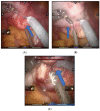
(A,B) Transverse incision in the anterior (posterior) wall of the uterus. Arrows point to the directions of incision.
Figure 3
Figure 3
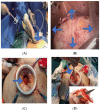
(A) Terlipressin was injected into the uterine body. (B) “Water cushion method” was used to isolate fibroids. (C) The fibroids were pulled with fibroid screws ("Kangji" brand fibroids screws). Arrows point to injection and pulling directions.
Figure 4
Figure 4
(A) The suture of the uterine wound. (B) 1-0 V-Loc absorbable barbed suture for continuous suture. Arrows point to suture direction.
Figure 5
Figure 5
(A) The extractor is inserted through an umbilical trocar. (B) The fibroids are placed in the specimen bag. (C) The incision is dilated with abdominal wall dilator. (D) The “peeling” method. (E) This is a fibroid removed through the umbilical hole. (F) The “bracelet” method. Arrows point to the location and method of fibroids removal and the fibroids removed.
Figure 5
Figure 5
(A) The extractor is inserted through an umbilical trocar. (B) The fibroids are placed in the specimen bag. (C) The incision is dilated with abdominal wall dilator. (D) The “peeling” method. (E) This is a fibroid removed through the umbilical hole. (F) The “bracelet” method. Arrows point to the location and method of fibroids removal and the fibroids removed.
Figure 6
Figure 6
(A) The patent for the fibroid extraction forceps. (B) The schematic diagram of myoma forceps used for endoscopy. In the picture: 1- telescopic tube, 2- duckbill clamp, 3- pulling device, 4- pressing handle, 5- adjusting gear, 7- rubber locking ring.
Figure 7
Figure 7
(A,B) Resection of myomatous with degeneration. Arrows point to the space between the degenerated fibroids and the myometrium.
Figure 8
Figure 8
(A,B) Resection of cervical fibroids. Arrows point to the location of the cervical fibroids.
Figure 9
Figure 9
(A,B) Resection of intravascular leiomyoma. Arrows point to the location of the intravascular leiomyoma.

Similar articles

Cited by

References

    1. Medikare V., Kandukuri L.R., Ananthapur V., Deenadayal M., Nallari P. The genetic bases of uterine fibroids; A review. J. Reprod. Infertil. 2011;12:181–191. - PMC - PubMed
    1. Munro M.G., Critchley H.O., Fraser I.S. The FIGO classification of causes of abnormal uterine bleeding: Malcolm G. Munro, Hilary O.D. Crithcley, Ian S. Fraser, for the FIGO Working Group on Menstrual Disorders. Int. J. Gynaecol. Obstet. 2011;113:1–2. doi: 10.1016/j.ijgo.2011.01.001. - DOI - PubMed
    1. Sandberg A.A. Updates on the cytogenetics and molecular genetics of bone and soft tissue tumors: Leiomyoma. Cancer Genet. Cytogenet. 2005;158:1–26. doi: 10.1016/j.cancergencyto.2004.08.025. - DOI - PubMed
    1. Bestel E., Donnez J. The potential of selective progesterone receptor modulators for the treatment of uterine fibroids. Expert Rev. Endocrinol. Metab. 2014;9:79–92. doi: 10.1586/17446651.2014.862495. - DOI - PubMed
    1. Islam M.S., Afrin S., Jones S.I., Segars J. Selective Progesterone Receptor Modulators-Mechanisms and Therapeutic Utility. Endocr. Rev. 2020;41:bnaa012. doi: 10.1210/endrev/bnaa012. - DOI - PMC - PubMed

LinkOut - more resources