Review
. 2022 May 27;12(11):1829.
doi: 10.3390/nano12111829.

Advances in Electrospun Hybrid Nanofibers for Biomedical Applications

Affiliations
Review

Advances in Electrospun Hybrid Nanofibers for Biomedical Applications

Viraj P Nirwan et al. Nanomaterials (Basel). .

Abstract

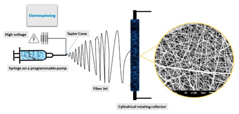
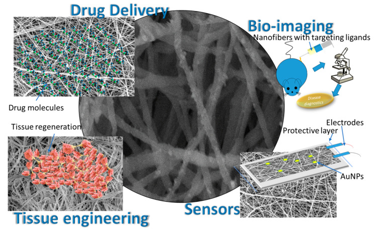
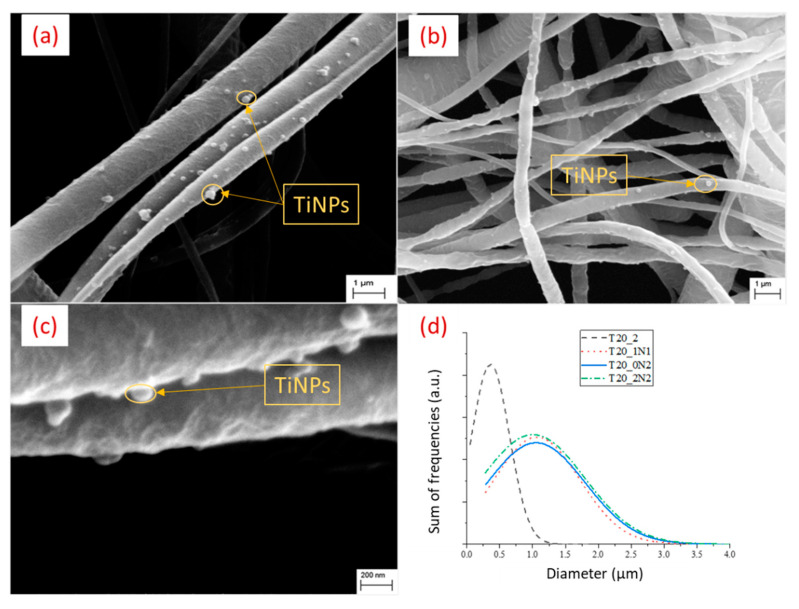
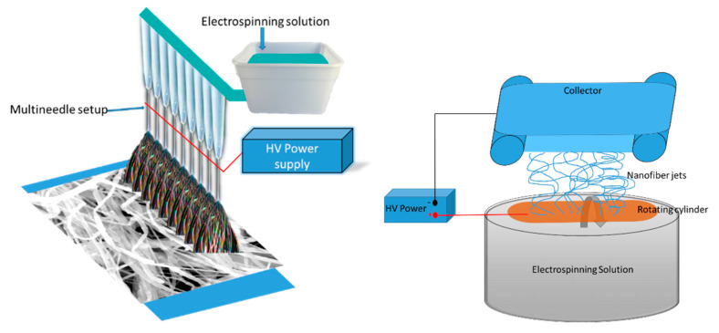
Electrospun hybrid nanofibers, based on functional agents immobilized in polymeric matrix, possess a unique combination of collective properties. These are beneficial for a wide range of applications, which include theranostics, filtration, catalysis, and tissue engineering, among others. The combination of functional agents in a nanofiber matrix offer accessibility to multifunctional nanocompartments with significantly improved mechanical, electrical, and chemical properties, along with better biocompatibility and biodegradability. This review summarizes recent work performed for the fabrication, characterization, and optimization of different hybrid nanofibers containing varieties of functional agents, such as laser ablated inorganic nanoparticles (NPs), which include, for instance, gold nanoparticles (Au NPs) and titanium nitride nanoparticles (TiNPs), perovskites, drugs, growth factors, and smart, inorganic polymers. Biocompatible and biodegradable polymers such as chitosan, cellulose, and polycaprolactone are very promising macromolecules as a nanofiber matrix for immobilizing such functional agents. The assimilation of such polymeric matrices with functional agents that possess wide varieties of characteristics require a modified approach towards electrospinning techniques such as coelectrospinning and template spinning. Additional focus within this review is devoted to the state of the art for the implementations of these approaches as viable options for the achievement of multifunctional hybrid nanofibers. Finally, recent advances and challenges, in particular, mass fabrication and prospects of hybrid nanofibers for tissue engineering and biomedical applications have been summarized.

Keywords: bone regeneration; drug delivery; electrospinning; functional agents; hybrid nanofibers; nanomedicine; nanoparticles; tissue engineering.


Conflict of interest statement

The authors declare no conflict of interest.

Figures

Figure 1
Figure 1
Schematic representation of simple electrospinning set up with rotating cylinder collector.
Figure 2
Figure 2
FESEM image of (a) TiO2 nanoparticles, (b) TiO2 nanoparticles with PVA/TiO2 nanofibers, (c) cross-sectional view of TiO2 nanoparticle, and (d) a cross-section of TiO2 nanoparticles with PVA/TiO2 nanofibers. Reproduced from Ref. [64].
Figure 3
Figure 3
Common biomedical applications of functionalized nanofibers.
Figure 4
Figure 4
SEM micrograph of AuNPs-functionalized Chitosan (PEO) nanofibers neutralized: (a) with 1M K2CO3 in 70% ethanol; (b) with 5M NaOH in methanol; (c) corresponding EDX spectroscopy graph showing presence of AuNPs after neutralizing with NaOH method; (d) FTIR spectra of nanofibers functionalized with AuNPs before neutralization (Ch-Au) and after neutralization with K2CO3 (Ch-Au/K2CO3) or NaOH (Ch-Au/NaOH). Adapted from Ref. [132].
Figure 5
Figure 5
Ligand-free TiN NPs-functionalized PCL (20% w/v) nanofibers with various concentrations of TiN NPs in electrospinning solutions: (a) 1 mL (0.15 mg L−1), T20_1N1; (b) 2 mL (0.15 mg L−1), T20_0N2; (c) 2 mL (0.45 mg L−1), T20_0N6; (d) statistical analysis of nanofibers’ diameter measured using ImageJ. Adapted from Ref. [157].
Figure 6
Figure 6
Biocompatible assays carried out on 3T3 fibroblasts immobilized on pristine PCL and TiN NPs-functionalized PCL scaffolds at various concentrations of NPs: (a) metabolic activity measured using the MTS assay; (b) proliferation using dsDNA assay; and (c) viability using live/dead assay. Tissue culture plastic (TCP) was chosen as a reference to provide the highest absorbance in MTS test. * refers to the statistical difference related to all other samples. No significant differences among scaffolds were observed in both cell proliferation and cell viability tests. In the statistics in (a), T8 and T10 in the above columns display statistical differences between groups T20_1N1 or T20_0N6, respectively. All assays show results as a mean and standard deviation. Reproduced from Ref. [157].
Figure 7
Figure 7
(a) Images of hDPSCs grown onto nanofiber scaffold for seven days demonstrate live hDPSCs forming colonies on PLCL surface confirmed by PKH26 red and DAPI staining (magnification ×400); (b) osteogenic differentiation of hDPSCs grown on PLCL stained by Alizarin Red S confirmed mineral deposits on PLCL fibers (Scale bar = 50 µm).
Figure 8
Figure 8
(a) Heat treatment of nanofiber under 160 °C. (b) Digital images of the area changes of the whole nanofiber containing gold nanorods (GNRs) upon irradiation of NIR light. (c) Change in area of the whole nanofiber containing GNRs as a function of cycles of temperature alternation upon the NIR irradiation. (d) Digital images of the area of the whole nanofiber without GNRs in the presence and absence of NIR light irradiation. Reproduced from Ref. [188].
Figure 9
Figure 9
Schematic representation of multiple needle and needleless electrospinning set up for generating higher yields.

Similar articles

Cited by

References

    1. Du L., Xu H., Zhang Y., Zou F. Electrospinning of polycaprolatone nanofibers with DMF additive: The effect of solution proprieties on jet perturbation and fiber morphologies. Fibers Polym. 2016;17:751–759. doi: 10.1007/s12221-016-6045-3. - DOI
    1. Kakoria A., Sinha-Ray S. A review on biopolymer-based fibers via electrospinning and solution blowing and their applications. Fibers. 2018;6:45. doi: 10.3390/fib6030045. - DOI
    1. Han D., Steckl A.J. Coaxial electrospinning formation of complex polymer fibers and their applications. ChemPlusChem. 2019;84:1453–1497. doi: 10.1002/cplu.201900281. - DOI - PubMed
    1. Patil J.V., Mali S.S., Kamble A.S., Hong C.K., Kim J.H., Patil P.S. Electrospinning: A versatile technique for making of 1D growth of nanostructured nanofibers and its applications: An experimental approach. Appl. Surf. Sci. 2017;423:641–674. doi: 10.1016/j.apsusc.2017.06.116. - DOI
    1. Kishan A.P., Cosgriff-Hernandez E.M. Recent advancements in electrospinning design for tissue engineering applications: A review. J. Biomed. Mater. Res. Part A. 2017;105:2892–2905. doi: 10.1002/jbm.a.36124. - DOI - PubMed

LinkOut - more resources