Review
. 2022 May 28;14(11):2189.
doi: 10.3390/polym14112189.

Functional Ultra-High Molecular Weight Polyethylene Composites for Ligament Reconstructions and Their Targeted Applications in the Restoration of the Anterior Cruciate Ligament

Affiliations
Review

Functional Ultra-High Molecular Weight Polyethylene Composites for Ligament Reconstructions and Their Targeted Applications in the Restoration of the Anterior Cruciate Ligament

Sonia B Wahed et al. Polymers (Basel). .

Abstract

The selection of biomaterials as biomedical implants is a significant challenge. Ultra-high molecular weight polyethylene (UHMWPE) and composites of such kind have been extensively used in medical implants, notably in the bearings of the hip, knee, and other joint prostheses, owing to its biocompatibility and high wear resistance. For the Anterior Cruciate Ligament (ACL) graft, synthetic UHMWPE is an ideal candidate due to its biocompatibility and extremely high tensile strength. However, significant problems are observed in UHMWPE based implants, such as wear debris and oxidative degradation. To resolve the issue of wear and to enhance the life of UHMWPE as an implant, in recent years, this field has witnessed numerous innovative methodologies such as biofunctionalization or high temperature melting of UHMWPE to enhance its toughness and strength. The surface functionalization/modification/treatment of UHMWPE is very challenging as it requires optimizing many variables, such as surface tension and wettability, active functional groups on the surface, irradiation, and protein immobilization to successfully improve the mechanical properties of UHMWPE and reduce or eliminate the wear or osteolysis of the UHMWPE implant. Despite these difficulties, several surface roughening, functionalization, and irradiation processing technologies have been developed and applied in the recent past. The basic research and direct industrial applications of such material improvement technology are very significant, as evidenced by the significant number of published papers and patents. However, the available literature on research methodology and techniques related to material property enhancement and protection from wear of UHMWPE is disseminated, and there is a lack of a comprehensive source for the research community to access information on the subject matter. Here we provide an overview of recent developments and core challenges in the surface modification/functionalization/irradiation of UHMWPE and apply these findings to the case study of UHMWPE for ACL repair.

Keywords: biofunctionalization; ligament; surface modification; synthetic graft; tendon; ultra-high molecular weight polyethylene.


Conflict of interest statement

The authors declare no conflict of interest.

Figures

Figure 1
Figure 1
Contrasting approaches for surface functionalization of UHMWPE.
Figure 2
Figure 2
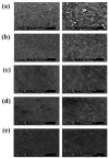
SEM images for (a) pristine UHMWPE fibers; (b) UHMWPE-PDA fibers; (c) UHMWPE-PDA-EDGE fibers; (d) UHMWPE − (PDA + EGDE) fibers; and micrographs of the water contact angle test. Reprinted with permission from [41].
Figure 3
Figure 3
(a) Pull out test of UHMWPE and modified fibers; (b) SEM images of untreated and treated UHMWPE; (i) UHMWPE fibers/rubber, (ii) UHMWPE–PDA fibers/rubber, (iii) UHMWPE–PDA–EGDE fibers/rubber, and (iv) UHMWPE– (PDA + EGDE) fibers/rubber. Reprinted with permission from [41].
Figure 4
Figure 4
Scanning electron microscopy of UHMWPE fibers with tensile fracture surface; (a) epoxy resin, (b) UHMWPE-epoxy resin, (c) UHMWPE-PDA-epoxy resin, and (d) UHMWPE-PDA-HMDA-epoxy resin. Reprinted with permission from [42].
Figure 5
Figure 5
(a) Wear rate comparison of conventional UHMWPE and UHMWPE-HA composite and control samples; (b) water contact angle and protein adsorption resistance results of untreated and treated UHMWPE; water flux and protein rejection of various membranes; (c) Water flux; and (d) HA and BSA rejection. Reprinted with permission from [44,45,73].
Figure 6
Figure 6
(a) Wear factors of untreated and treated UHMWPE after cold plasma treatment, reprinted with permission from [89]; (b) osteoblast cell adhesion to untreated and cold plasma treated UHMWPE, reprinted with permission from [89]; (c) a schematic diagram of cold atmospheric plasma.
Figure 7
Figure 7
L929 cell attachment on untreated and treated UHMWPE. Reprinted with permission from [93].
Figure 8
Figure 8
(a) Plasma-assisted chemical vapor deposition (PACVD) coating of UHMWPE fabric; (b) introduction of additional groups into UHMWPE before and after treatment, reprinted with permission from [34]; (c) (i) friction coefficient of friction of untreated and treated UHMWPE; (ii) scratch penetration vs. load graph, reprinted with permission from [34]; (d) differentiation of PBMNCs to osteoclast a. Untreated UHMWPE, b. HN-1 min, c. O2-1 min, and d. HN-2 min do not display presence of osteoclasts, whereas e. O2-2 min displays the presence of multinucleated giant osteoclasts on its surface (indicated with arrows). Reprinted with permission from [33].
Figure 9
Figure 9
(a) Plasma treatment of UHMWPE fabric in an electron-cyclotron resonance (ECR) plasma reactor system. (b) Dielectric barrier discharge (DBD) plasma treatment of polyethylene fabric. (c) Impact of plasma treatment time on tensile strength of modified DBD-chitosan treatment UHMWPE fibers. Reprinted with permission from [47]. (d) Tensile strength of untreated and UV treated UHMWPE. Reprinted with permission from [18]. (e) The influence of degree of graft on tensile strength. Reprinted with permission from [99].
Figure 10
Figure 10
(a) Water contact angle of UHMWPE before and after protein incubation. Reprinted with permission from [19]. (b) AFM images of UHMWPE with or without protein incubation; (a) pure UHMWPE; (b) after incubation in BSA solution; (c) after incubation in NaHA solution; (d) after incubation in both solutions. Reprinted with permission from [19]. (c) Water contact angle of treated and untreated UHMWPE after surface treatment. Reprinted with permission from [145]. (d) The force displacement curve of three-point bending test for (i) freeze-dried collagen hybrid; (ii) collagen-HAp hybrid. Reprinted with permission from [145]. (e) Antifouling properties of different membranes (M1, M2, M3, M4, and M5): (i) variation of time-dependent flux over three periods with bovine serum albumin (BSA) as a pollutant; (ii) values of FRR with BSA as a pollutant. Reprinted with permission from [70]. (f) Contact angle vs. drop age curves for the original and modified PE porous membranes: (i) untreated PE membrane; (ii) PE-polydopamine composite membrane; (cf) PE/dopamine-heparin composite membrane; (b) impact of heparin immobilization period for PE membranes on water flux. Reprinted with permission from [65].
Figure 11
Figure 11
Unmodified and modified PE porous membrane surface platelet morphology: (a) initial PE membrane, (b) PE/dopamine composite membrane, (c,d) PE/dopamine-heparin composite membrane, (ce). Reprinted with permission from [65].
Figure 12
Figure 12
(a) The bar graph represents the coefficient of friction vs. protein concentration for all tested protein solutions at different sliding speed: (i) 10 mm and (ii) 50 mm. Reprinted with permission from [153]. (b) Live (green) and dead (red) cells on PE/PCL a,c, and PE/PCL/BG composites b,d after one a,b and seven c,d days of culture [154].
Figure 13
Figure 13
(a) Bioactivity of bioglass; (b) schematic preparation of SF/VEGF coating [37]; (c) morphology of different groups of UHMWPE [37].

References

    1. Gobbi S.J., Gobbi J.V., Rocha Y. Requirements for Selection/Development of a Biomaterial. Biomed. J. Sci. Tech. Res. 2019;14:1–6. doi: 10.26717/BJSTR.2019.14.002554. - DOI
    1. Geetha M., Singh A.K., Asokamani R., Gogia A.K. Ti Based Biomaterials, the Ultimate Choice for Orthopaedic Implants—A Review. Prog. Mater. Sci. 2009;54:397–425. doi: 10.1016/j.pmatsci.2008.06.004. - DOI
    1. Kurtz S.M., Muratoglu O.K., Evans M., Edidin A.A. Advances in the Processing, Sterilization, and Crosslinking of Ultra-High Molecular Weight Polyethylene for Total Joint Arthroplasty. Biomaterials. 1999;20:1659–1688. doi: 10.1016/S0142-9612(99)00053-8. - DOI - PubMed
    1. Oosterom R., Ahmed T.J., Poulis J.A., Bersee H.E.N. Adhesion Performance of UHMWPE after Different Surface Modification Techniques. Med. Eng. Phys. 2006;28:323–330. doi: 10.1016/j.medengphy.2005.07.009. - DOI - PubMed
    1. Diabb Zavala J.M., Leija Gutiérrez H.M., Segura-Cárdenas E., Mamidi N., Morales-Avalos R., Villela-Castrejón J., Elías-Zúñiga A. Manufacture and Mechanical Properties of Knee Implants Using SWCNTs/UHMWPE Composites. J. Mech. Behav. Biomed. Mater. 2021;120:104554. doi: 10.1016/j.jmbbm.2021.104554. - DOI - PubMed

LinkOut - more resources