. 2022 May 26;14(11):2211.
doi: 10.3390/nu14112211.

The Neuroprotective Effects of Spray-Dried Porcine Plasma Supplementation Involve the Microbiota-Gut-Brain Axis

Affiliations

The Neuroprotective Effects of Spray-Dried Porcine Plasma Supplementation Involve the Microbiota-Gut-Brain Axis

Cristina Rosell-Cardona et al. Nutrients. .

Abstract

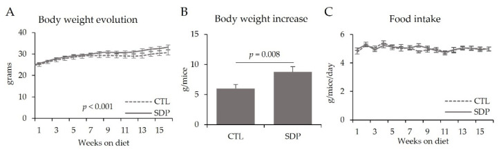
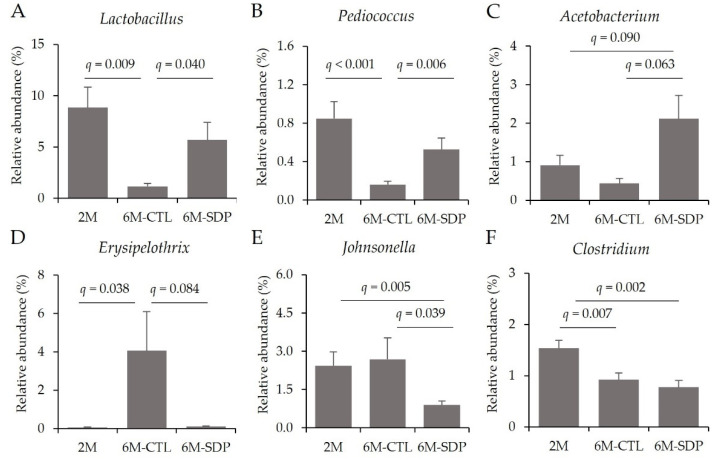
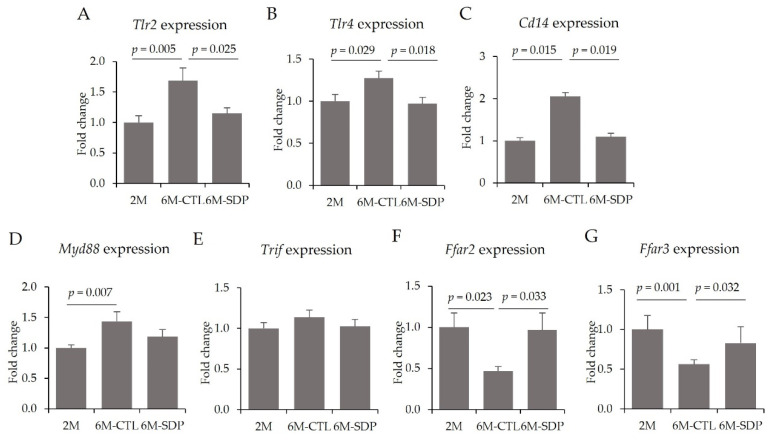
Dietary supplementation with spray-dried porcine plasma (SDP) reduces the Alzheimer’s disease (AD) hallmarks in SAMP8 mice. Since gut microbiota can play a critical role in the AD progression, we have studied if the neuroprotective effects of SDP involve the microbiota−gut−brain axis. Experiments were performed on two-month-old SAMP8 mice fed a standard diet and on six-month-old SAMP8 mice fed a control diet or an 8% SDP supplemented diet for four months. Senescence impaired short- and long-term memory, reduced cortical brain-derived neurotrophic factor (BDNF) abundance, increased interleukin (Il)-1β, Il-6, and Toll-like receptor 2 (Tlr2) expression, and reduced transforming growth factor β (Tgf-β) expression and IL-10 concentration (all p < 0.05) and these effects were mitigated by SDP (all p < 0.05). Aging also increased pro-inflammatory cytokines in serum and colon (all p < 0.05). SDP attenuated both colonic and systemic inflammation in aged mice (all p < 0.05). SDP induced the proliferation of health-promoting bacteria, such as Lactobacillus and Pediococcus, while reducing the abundance of inflammation-associated bacteria, such as Johnsonella and Erysipelothrix (both q < 0.1). In conclusion, SDP has mucosal and systemic anti-inflammatory effects as well as neuroprotective properties in senescent mice; these effects are well correlated with SDP promotion of the abundance of probiotic species, which indicates that the gut−brain axis could be involved in the peripheral effects of SDP supplementation.

Keywords: Alzheimer’s disease; aging; dietary supplementation; microbiota; spray-dried porcine plasma.


Conflict of interest statement

C.R.-C., C.A., C.G.-F., M.P., A.P.-B., M.M. and L.M. have no conflict of interests. J.P. is employed by APC-Europe S.L.U. The funding sponsors had no role in the design of the study; in the collection, analysis, or interpretation of data; in the writing of the manuscript, and in the decision to publish the results.

Figures

Figure 1
Figure 1
Body weight evolution (A), body weight increase (B) and food intake evolution (C) of SAMP8 mice during the experimental period. Results are expressed as mean ± SEM (n = 11–12 mice/group). Statistics: t-Student test.
Figure 2
Figure 2
Open field test (OFT). Representative tracking maps of 2M, 6M-CTL, and 6M-SDP groups (A), locomotor activity (B), exploratory activity (C) and percentage of time in the border zone (D). Results are expressed as mean ± SEM (n = 11–12 mice/group). Statistics: ANOVA (Fisher multiple comparison test), and Kruskal–Wallis test.
Figure 3
Figure 3
Novel object recognition test (NORT). Explorative index (A), object preference (B), short-term memory (C), long-term memory (D). Results are expressed as mean ± SEM (n = 11–12 mice/group). Statistics: ANOVA (Fisher multiple comparison test).
Figure 4
Figure 4
Abundance of BDNF precursor (pro-BDNF) (A) and mature BDNF (m-BDNF) (B). Results are expressed as mean ± SEM (n = 5−6 mice/group). Statistics: ANOVA (Fisher multiple comparison test), and chi-square test.
Figure 5
Figure 5
Neuroinflammation in cortical tissue. Gene expression of Il-1β (A) and Il-6 (B), protein abundance of p65-NF-κB respect to total NF-κB (C), gene expression of Tgf-β (D), and concentration of IL-10 (E). Results are expressed as mean ± SEM (n = 8–9 mice/group). Statistics: ANOVA (Fisher multiple comparison test). Il, interleukin; NF-κB, nuclear factor kappa-light-chain-enhancer of activated B cells; Tgf-β, transforming growth factor-beta.
Figure 6
Figure 6
TLR signaling pathway in cortical tissue. Gene expression of Tlr2 (A), Tlr4 (B), Cd14 (C), Myd88 (D), Trif (E), and concentration of LPS (F). Results are expressed as mean ± SEM (n = 6–9 mice/group). Statistics: ANOVA (Fisher multiple comparison test). Cd14, cluster of differentiation 14; LPS, lipopolysaccharide; Myd88, myeloid differentiation factor 88; Tlr, toll-like receptor; Trif, toll-like receptor adaptor molecule 1.
Figure 7
Figure 7
Systemic inflammation. Concentrations of LPS (A), IL-1β (B), TNF-α (C), and IL-10 (D). Results are expressed as mean ± SEM (n = 8–9 mice/group). Statistics: ANOVA (Fisher multiple comparison test). IL, interleukin; LPS, lipopolysaccharide; TNF-α, tumor necrosis factor-alpha.
Figure 8
Figure 8
Microbial Shannon’s index (A), species richness (B), ratio between Firmicutes/Bacteroidetes (F/B) (C), and bacterial composition at the phylum level in the 2M group (D), 6M-CTL group (E), and 6M-SDP group (F). Results are expressed as mean ± SEM and as percentages (n = 10–11 mice/group). Statistics: ANOVA (Benjamini and Hochberg multiple comparison test).
Figure 9
Figure 9
Fecal microbial composition at the genus level of the Firmicutes phylum. Relative abundance of Lactobacillus (A), Pediococcus (B), Acetobacterium (C), Erysipelothrix (D), Johnsonella (E), and Clostridium (F). Results are expressed as mean ± SEM (n = 10–11 mice/group). Statistics: ANOVA (False discovery rate, Benjamini and Hochberg multiple comparison test).
Figure 10
Figure 10
Fecal microbial composition at the genus level of the Bacteroidetes phylum. Relative abundance of Bacteroides (A), Prevotella (B) Odoribacter (C), and Parabacteroides (D). Results are expressed as mean ± SEM (n = 10–11 mice/group). Statistics: ANOVA (False discovery rate, Benjamini and Hochberg multiple comparison test).
Figure 11
Figure 11
Gene expression of the TLR signaling pathway and SCFA receptors in colon mucosa. Tlr2 (A), Tlr4 (B), Cd14 (C), Myd88 (D), Trif (E), Ffar2 (F), and Ffar3 (G). Results are expressed as mean ± SEM (n = 9–11 mice/group). Statistics: ANOVA (Fisher multiple comparison test). Cd14, cluster of differentiation 14; Ffar2, free fatty acid receptor 2 (GPR43); Ffar3, free fatty acid receptor 3 (GPR41); Myd88, myeloid differentiation factor 88; Tlr, toll-like receptor; Trif, toll-like receptor adaptor molecule 1.
Figure 12
Figure 12
Inflammation in colon tissue. Gene expression of Il-1β (A), Il-6 (B), Tnf-α (C), Cd25 (D), F4/80 (E), and Itgae (F). Results are expressed as mean ± SEM (n = 9−11 mice/group). Statistics: ANOVA (Fisher multiple comparison test) and Kruskal–Wallis test. Cd25, cluster of differentiation 25; Itgae, integrin alpha e; Il, interleukin; Tnf-α, tumor necrosis factor alpha.
Figure 13
Figure 13
Intestinal barrier. Gene expression of Tff3 (A), Muc2 (B), and Occludin (C). Results are expressed as mean ± SEM (n = 9−11 mice/group). Statistics: ANOVA (Fisher multiple comparison test). Muc2, mucin 2; Tff3, trefoil factor 3.
Figure 14
Figure 14
Schematic integrative representation of the effects of SDP supplementation on the gut−brain axis in an AD mouse model. Dietary supplementation with SDP promotes the growth of probiotic genera and reduces the levels of pathogenic genera. This is accompanied by a reduction in the intestinal permeability and local and systemic immune and inflammatory responses. These effects culminate in a reduction in neuroinflammation and an amelioration of the cognitive decline. AD, Alzheimer’s disease; GPCR, G protein-coupled receptors; SDP, spray-dried porcine plasma; TLR, toll-like receptor.

Similar articles

Cited by

References

    1. Lane C.A., Hardy J., Schott J.M. Alzheimer’s disease. Eur. J. Neurol. 2018;25:59–70. doi: 10.1111/ene.13439. - DOI - PubMed
    1. Calsolaro V., Edison P. Neuroinflammation in Alzheimer’s disease: Current evidence and future directions. Alzheimer’s Dement. 2016;12:719–732. doi: 10.1016/j.jalz.2016.02.010. - DOI - PubMed
    1. Soria-Lopez J.A., González H.M., Léger G.C. Alzheimer’s disease. In: DeKosky S.T., Asthana S., editors. Handbook of Clinical Neurology. 3rd ed. Volume 167. Elsevier; Boston, MA, USA: 2019. pp. 231–255. - DOI - PubMed
    1. Hou Y., Dan X., Babbar M., Wei Y., Hasselbalch S.G., Croteau D.L., Bohr V.A. Ageing as a risk factor for neurodegenerative disease. Nat. Rev. Neurol. 2019;15:565–581. doi: 10.1038/s41582-019-0244-7. - DOI - PubMed
    1. Breijyeh Z., Karaman R. Comprehensive Review on Alzheimer’s Disease: Causes and Treatment. Molecules. 2020;25:5789. doi: 10.3390/molecules25245789. - DOI - PMC - PubMed

Substances

LinkOut - more resources