. 2022 Jun 3;27(11):3597.
doi: 10.3390/molecules27113597.

Synthesis and Anticancer Properties of New 3-Methylidene-1-sulfonyl-2,3-dihydroquinolin-4(1 H)-ones

Affiliations

Synthesis and Anticancer Properties of New 3-Methylidene-1-sulfonyl-2,3-dihydroquinolin-4(1 H)-ones

Agata Jaskulska et al. Molecules. .

Abstract

Quinolinones have been known for a long time as broad-spectrum synthetic antibiotics. More recently, the anticancer potential of this group of compounds has been investigated. Following this direction, we obtained a small library of 3-methylidene-1-sulfonyl-2,3-dihydroquinolin-4(1H)-ones with various substituents at positions 1, 2, 6, and 7 of the quinolinone ring system. The cytotoxic activity of the synthesized analogs was tested in the MTT assay on two cancer cell lines in order to determine the structure-activity relationship. All compounds produced high cytotoxic effects in MCF-7, and even higher in HL-60 cells. 2-Ethyl-3-methylidene-1-phenylsulfonyl-2,3-dihydroquinolin-4(1H)-one, which was over 5-fold more cytotoxic for HL-60 than for normal HUVEC cells, was selected for further tests. This analog was shown to inhibit proliferation and induce DNA damage and apoptosis in HL-60 cells.

Keywords: Horner–Wadsworth–Emmons olefination; apoptosis; cytotoxicity; structure–activity relationship.


Conflict of interest statement

The authors declare no conflict of interest.

Figures

Figure 1
Figure 1
Chemical structure of quinoline, 4-quinolinones, and some diversely substituted analogs with quinolone skeleton.
Scheme 1
Scheme 1
Synthesis of 3-methylidene-1-sulfonyl-2,3-dihydroquinolin-4(1H)-ones 5a-t.
Figure 2
Figure 2
Structure of the selected compound 5a (a). Metabolic activity of (b) HL-60 and HUVEC cells treated with various concentrations of 5a for 48 h; (c) HL-60 cells treated with various concentration of 5a for 24 h, measured by MTT assay.
Figure 3
Figure 3
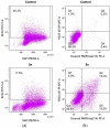
Representative scattered blots of multiparameter flow cytometry analysis of cell proliferation, DNA damage and apoptosis in HL-60 cells treated with 5a (1.8µM) for 24 h. Panel (a): DAPI vs. BrdU PerCP-Cy.5.5 staining profile. BrdU positive cells are in the inside frame. Panel (b): cleaved PARP (Asp214) PE vs. H2AX (pS139) Alexa profile. Squares Q1 + Q2 represent H2AX (pS139) Alexa positive cells, squares Q2 + Q4 represent cleaved PARP (Asp214) PE positive cells, whereas square Q3 represents H2AX (pS139) Alexa and cleaved PARP (Asp214) PE negative cells.
Figure 4
Figure 4
Quantitative analysis of inhibition of cell proliferation (BrdU test); (a) induction of apoptosis (PARP test) (b) and induction of DNA damage (H2AX test) (c) by 5a (1.8 µM) in HL-60 cells. Data are presented as mean ± SEM of three independent experiments. Statistical significance was assessed using one-way ANOVA and a post-hoc multiple comparison Student–Newman–Keuls test. *** p < 0.001 vs. control.
Figure 5
Figure 5
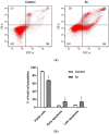
Induction of apoptosis in HL-60 cells treated with 5a (1.8 µM) for 24 h: (a) Representative scattered blots of flow cytometry analysis of apoptosis by double-staining with FITC-Annexin-V and PI. The percentage of viable cells is shown in quadrant Q3. Quadrant Q4 indicates the percentage of early apoptotic cells (Annexin V-positive cells), whereas quadrant Q2 shows the percentage of late apoptotic/death cells (Annexin V-and PI-positive cells). (b) Quantitative analysis of apoptotic cell death by Annexin V and PI assay. Data are presented as mean ±SEM of three independent experiments. Statistical significance was assessed using Student’s t test. *** p < 0.001 vs. control.
Figure 6
Figure 6
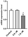
Real-time PCR analysis of ABCB1 mRNA levels in HL-60 cells treated with 5a for 24 h. Data are presented as mean ± SEM of three independent experiments. Statistical significance was assessed using one-way ANOVA and a post-hoc multiple comparison Student–Newman–Keuls test. * p < 0.05 vs. control.

References

    1. Shiro T., Fukaya T., Tobe M. The chemistry and biological activity of heterocycle-fused quinolinone derivatives: A review. Eur. J. Med. Chem. 2015;97:397–408. doi: 10.1016/j.ejmech.2014.12.004. - DOI - PubMed
    1. Beniddir M.A., Le Borgne E., Iorga B.I., Loaec N., Lozach O., Meijer L., Awang K., Litaudon M. Acridone alkaloids from Glycosmis chlorosperma as DYRK1A inhibitors. J. Nat. Prod. 2014;77:1117–1122. doi: 10.1021/np400856h. - DOI - PubMed
    1. Cretton S., Dorsaz S., Azzollini A., Favre-Godal Q., Marcourt L., Ebrahimi S.N., Voinesco F., Michellod E., Sanglard D., Gindro K., et al. Antifungal Quinoline Alkaloids from Waltheria indica. J. Nat. Prod. 2016;79:300–307. doi: 10.1021/acs.jnatprod.5b00896. - DOI - PubMed
    1. Emmerson A.M., Jones A.M. The quinolones: Decades of development and use. J. Antimicrob. Chemother. 2003;51:13–20. doi: 10.1093/jac/dkg208. - DOI - PubMed
    1. Kahriman N., Iskender N.Y., Yucel M., Yayli N., Demir E., Demirbag Z. Microwave-assisted synthesis of 1,3′-diaza-flavanone/flavone and their alkyl derivatives with antimicrobial activity. J. Heterocycl. Chem. 2012;49:71–79. doi: 10.1002/jhet.800. - DOI

LinkOut - more resources