. 2022 Jun 6;22(11):4297.
doi: 10.3390/s22114297.

BrainGAN: Brain MRI Image Generation and Classification Framework Using GAN Architectures and CNN Models

Affiliations

BrainGAN: Brain MRI Image Generation and Classification Framework Using GAN Architectures and CNN Models

Halima Hamid N Alrashedy et al. Sensors (Basel). .

Abstract

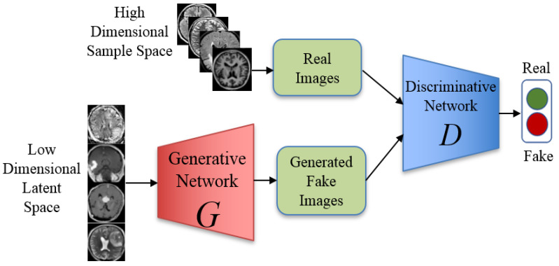
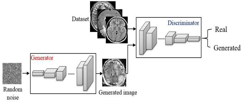
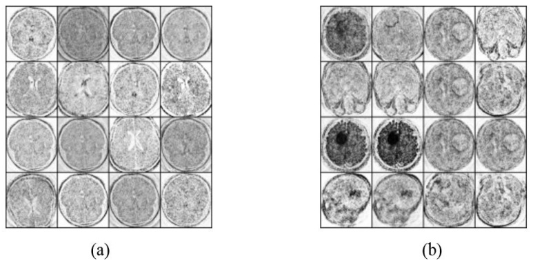
Deep learning models have been used in several domains, however, adjusting is still required to be applied in sensitive areas such as medical imaging. As the use of technology in the medical domain is needed because of the time limit, the level of accuracy assures trustworthiness. Because of privacy concerns, machine learning applications in the medical field are unable to use medical data. For example, the lack of brain MRI images makes it difficult to classify brain tumors using image-based classification. The solution to this challenge was achieved through the application of Generative Adversarial Network (GAN)-based augmentation techniques. Deep Convolutional GAN (DCGAN) and Vanilla GAN are two examples of GAN architectures used for image generation. In this paper, a framework, denoted as BrainGAN, for generating and classifying brain MRI images using GAN architectures and deep learning models was proposed. Consequently, this study proposed an automatic way to check that generated images are satisfactory. It uses three models: CNN, MobileNetV2, and ResNet152V2. Training the deep transfer models with images made by Vanilla GAN and DCGAN, and then evaluating their performance on a test set composed of real brain MRI images. From the results of the experiment, it was found that the ResNet152V2 model outperformed the other two models. The ResNet152V2 achieved 99.09% accuracy, 99.12% precision, 99.08% recall, 99.51% area under the curve (AUC), and 0.196 loss based on the brain MRI images generated by DCGAN architecture.

Keywords: DCGANs; brain MRI images; deep learning; image classification; image generation; vanilla GANs.


Conflict of interest statement

The authors declare no conflict of interest.

Figures

Figure 1
Figure 1
Our proposed BrainGAN framework starting with Brain MRI dataset real images, generating images using DCGAN and Vanilla GAN, deep learning models CNN, MobileNetV2, and ResNet152V2, and finally the testing & validation.
Figure 2
Figure 2
The Pseudo-code of the proposed BrainGAN framework.
Figure 3
Figure 3
MRI scan images for two classes (a) No tumor samples images, and (b) Tumor sample images.
Figure 4
Figure 4
The proposed architecture of the Vanilla GANs to generate MRI images.
Figure 5
Figure 5
The proposed architecture of the DCGANs to generate MRI images.
Figure 6
Figure 6
MRI scan images that are generated by applying Vanilla GANs for (a) Vanilla GANs no tumor images, and (b) Vanilla GANs tumor images.
Figure 7
Figure 7
MRI scan images that are generated by applying DCGANs for (a) DCGANs no tumor images, and (b) DCGANs tumor images.
Figure 8
Figure 8
Confusion matrix for the proposed CNN model: (a) using Vanilla GAN image generated; (b) using DCGAN image generated.
Figure 9
Figure 9
Loss, AUC, precision, recall, and accuracy between the training and validation phases with the number of epochs for the CNN model using DCGAN image generated.
Figure 10
Figure 10
Confusion matrix for the proposed MobileNetV2 model: (a) using Vanilla GAN image generated; (b) using DCGAN image generated.
Figure 11
Figure 11
Loss, AUC, precision, recall, and accuracy between the training and validation phases with the number of epochs for the MobileNetV2 model using DCGAN image generated.
Figure 12
Figure 12
Confusion matrix for the proposed ResNet152V2 model: (a) using Vanilla GAN image generated; (b) using DCGAN image generated.
Figure 13
Figure 13
Loss, AUC, precision, recall, and accuracy between the training and validation phases with the number of epochs for the ResNet152V2 model using DCGAN image generated.
Figure 14
Figure 14
Loss measures for the CNN, MobileNetV2, ResNet152V2 models using Vanilla GAN and DCGAN image generated.
Figure 15
Figure 15
Accuracy, precision, recall, and area under the curve (AUC) measures for the proposed CNN, MobileNetV2, ResNet152V2 models using Vanilla GAN and DCGAN image generated.
Figure 16
Figure 16
Accuracy performance metrics comparison between our proposed models and previous studies [11,18,20].
Figure 17
Figure 17
Precision performance metrics comparison between our proposed models and previous studies [18].
Figure 18
Figure 18
Recall performance metrics comparison between our proposed models and previous studies [18].

References

    1. Goodfellow I., Pouget-Abadie J., Mirza M., Xu B., Warde-Farley D., Ozair S., Courville A., Bengio Y. Generative adversarial nets. Adv. Neural Inf. Process. Syst. 2014;3:2672–2680.
    1. Kumar S., Dabas C., Godara S. Classification of brain MRI tumor images: A hybrid approach. Procedia Comput. Sci. 2017;122:510–517. doi: 10.1016/j.procs.2017.11.400. - DOI
    1. Gab Allah A.M., Sarhan A.M., Elshennawy N.M. Classification of Brain MRI Tumor Images Based on Deep Learning PGGAN Augmentation. Diagnostics. 2021;11:2343. doi: 10.3390/diagnostics11122343. - DOI - PMC - PubMed
    1. Wu D., Rice C.M., Wang X. Cancer bioinformatics: A new approach to systems clinical medicine. BMC Bioinform. 2012;13:S7. doi: 10.1186/1471-2105-13-71. - DOI - PMC - PubMed
    1. Brain MRI Segmentation. 2019. [(accessed on 5 March 2022)]. Available online: https://www.kaggle.com/mateuszbuda/lgg-mri-segmentation.