. 2022 May 24:10:892015.
doi: 10.3389/fbioe.2022.892015. eCollection 2022.

Lycorine Inhibits Hypertrophic Scar Formation by Inducing ROS-Mediated Apoptosis

Affiliations

Lycorine Inhibits Hypertrophic Scar Formation by Inducing ROS-Mediated Apoptosis

Yunxian Dong et al. Front Bioeng Biotechnol. .

Abstract

Background: Hypertrophic scar (HS) is a fibrotic cutaneous disease with few effective therapies. Lycorine is a drug with pro-apoptotic ability and anti-fibrosis potential. This study aimed to test whether lycorine could trigger the apoptosis of hypertrophic scar fibroblasts (HSFs) to inhibit HS formation. Methods: The proapoptotic and anti-fibrosis effects of lycorine on the viability and apoptosis of human primary HSFs and their reactive oxygen species (ROS) production as well as a rabbit ear model of HS were determined by CCK-8, flow cytometry, Western blot, immunofluorescence, transwell migration, collagen gel contraction assays. Results: Lycorine treatment selectively decreased the viability of HSFs, and induced their apoptosis, but not normal fibroblasts (NFs). Lycorine treatment increased the relative levels of Bax and cleaved PARP expression, cytochrome C cytoplasm translocation, but decreased Bcl-2, caspase-3 and caspase-9 expression, the mitochondrial membrane potential (MMP) in HSFs. Lycorine inhibited the migration and contraction of HSFs, and reduced the expression of collagen I, collagen III and α-SMA. Mechanistically, lycorine treatment stimulated high levels of ROS production, leading to apoptosis of HSFs while treatment with NAC, a ROS inhibitor, significantly mitigated or abrogated the pro-apoptotic and antifibrotic activity of lycorine in HSFs. Moreover, lycorine treatment mitigated the severity of HS in rabbit ears by inducing fibroblast apoptosis. Conclusion: These results indicate that lycorine has a potent anti-fibrotic activity and is a potential drug for intervention of HS.

Keywords: apoptosis; fibrosis; hypertrophic scar; lycorine; reactive oxygen species.


Conflict of interest statement

The authors declare that the research was conducted in the absence of any commercial or financial relationships that could be construed as a potential conflict of interest.

Figures
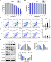
FIGURE 1
FIGURE 1
Lycorine reduces the viability of HSFs by triggering their apoptosis. HSFs and NFs were treated with vehicle or lycorine at indicated concentrations. At 12, 24 and 48 h later, cell viability (n = 5) of HSFs (A) and NFs (B) were determined by CCK-8 assay. Flow cytometric analysis of the frequency of apoptotic HSFs (C) and NFs (D) at 24 h post lycorine treatment (n = 4). (E) At 24 h post lycorine treatment, the relative levels of Bax, Bcl-2, caspase 9, caspase3, PARP and cleaved PARP (c-PARP) in HSFs were examined by Western blot (n = 3). Data are mean ± standard error of the mean. *p < 0.05, **p < 0.01, ***p < 0.001.
FIGURE 2
FIGURE 2
Lycorine induced mitochondrial apoptosis of HSFs. (A) HSFs were treated with vehicle or lycorine for 24 h, and the mitochondrial membrane potential (MMP) was analyzed by fluorescent imaging after staining the cells with JC-1 (n = 3). Scale bar: 200 μM. (B) At 24 h post lycorine treatment, the relative levels of Cytochrome C (cyt-C) in both the cytoplasm and mitochondria were examined by Western blot (n = 3). (C) At 24 h post lycorine treatment, the relative levels of caspase 8, Fas, Fasl to the control GAPDH expression in NFs were examined by Western blot (n = 3). Data are mean ± standard error of the mean.
FIGURE 3
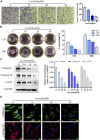
FIGURE 3
Lycorine inhibits the migration and contraction of HSFs by reducing their production of fibrotic factors. HSFs were treated with vehicle or lycorine at indicated concentrations. (A) Transwell migration assays indicated that lycorine treatment for 24 h inhibited the migration of HSFs (n = 3). Scale bar = 200 mm. (B) HSFs were mixed with rat tail collagen, and the collagen gesl were imaged at 12 and 24 h later (n = 3). (C) At 24 h post lycorine treatment, the relative levels of a-smooth muscle actin (a-SMA), collagen I, and collagen III expression were determined by Western blot (n = 3) and (D) the levels of collagen I, collagen III expression were analyzed by immunofluorescence (n = 4), Scale bar = 20 μM. Data are mean ± standard error of the mean. *p < 0.05, **p < 0.01, ***p < 0.001.
FIGURE 4
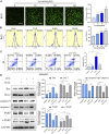
FIGURE 4
Lycorine stimulates high levels of reactive oxygen species (ROS) production, leading to the apoptosis of HSFs. (A–B) HSFs were treated with vehicle or lycorine at indicated concentrations. The levels of intracellular ROS were analyzed by (A) immunofluorescence (n = 4, Scale bar = 400 μM) and (B) flow cytometry (n = 4). (C)Flow cytometric analysis of the percentages of apoptotic HSFs following treatment with vehicle (control) or 40 μM lycorine in the presence or absence of 100 μM NAC for 24 h (n = 4). (D) The relative levels of a-smooth muscle actin (a-SMA), collagen I, and collagen III expression in the different groups of cells were analyzed by Western blot (n = 3). Data are mean ± standard error of the mean. *p < 0.05, **p < 0.01, ***p < 0.001.
FIGURE 5
FIGURE 5
High levels of ROS induced by lycorine alters the mitochondrial membrane potential (MMP) and cytochrome C (cyt-C) translocation. HSFs were treated with vehicle (control) or 40 μM lycorine in the presence or absence of 100 μM NAC for 24 h. (A) The MMP was analyzed by fluorescence microscopy after staining with JC-1 (n = 3). Scale bar: 400 μM. (B) The levels of Cytochrome C (cyt-C) in the both cytoplasm and mitochondria were examined by Western blot (n = 3). Data are mean ± standard error of the mean. *p < 0.05, **p < 0.01, ***p < 0.001.
FIGURE 6
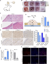
FIGURE 6
Lycorine treatment attenuates the severity of hypertrophic scar (HS) in vivo. At 14 days post surgery in rabbit ears, the rabbits were randomized and injected subcutaneously with vehicle DMSO or lycorine into each rabbit’s ear. (A) Schematic diagram of a rabbit ear HS model. (B) The wounded ears and scar formation at the indicated time points. (C) HE staining of scar tissue sections at 28 days post surgery (n = 4). “←→” indicates the scar thickness. Scale bar = 100 mm. (D) The analysis of SEI at 28 days post surgery. SEI > 1 indicates HS (n = 4). (E) Immunohistochemistry analysis of a-smooth muscle actin (a-SMA) , collagen I and Collagen III expression (n = 4), Scale bar = 100 mm. (F) The relative levels of Bax and Bcl-2 mRNA transcripts in rabbit ear scar tissues at 28 days post surgery (n = 4). (G) Immunofluorescence analysis of caspase 9 expression in rabbit ear scar tissues (n = 4). Data are mean ± standard error of the mean. *p < 0.05, **p < 0.01, ***p < 0.001.
FIGURE 7
FIGURE 7
A Schematic diagram for ROS-mediated mitochondrial apoptosis of lycorine in hypertrophic scar fibroblasts (HSFs).

Similar articles

Cited by

References

    1. Bendaif H., Melhaoui A., Ramdani M., Elmsellem H., Douez C., El Ouadi Y. (2018). Antibacterial Activity and Virtual Screening by Molecular Docking of Lycorine from Pancratium Foetidum Pom (Moroccan Endemic Amaryllidaceae). Microb. Pathog. 115, 138–145. 10.1016/j.micpath.2017.12.037 - DOI - PubMed
    1. Cadenas S. (2018). Mitochondrial Uncoupling, ROS Generation and Cardioprotection. Biochim. Biophys. Acta (BBA) - Bioenerg. 1859 (9), 940–950. 10.1016/j.bbabio.2018.05.019 - DOI - PubMed
    1. Cao Z., Yang P., Zhou Q. (2013). Multiple Biological Functions and Pharmacological Effects of Lycorine. Sci. China Chem. 56 (10), 1382–1391. 10.1007/s11426-013-4967-9 - DOI - PMC - PubMed
    1. Chen M., Liu X., Hu B., Fan Z., Song Y., Wei H., et al. (2018). Rabbit Hemorrhagic Disease Virus Non-structural Protein 6 Induces Apoptosis in Rabbit Kidney Cells. Front. Microbiol. 9, 3308. 10.3389/fmicb.2018.03308 - DOI - PMC - PubMed
    1. Danial N. N. (2007). BCL-2 Family Proteins: Critical Checkpoints of Apoptotic Cell Death. Clin. Cancer Res. 13 (24), 7254–7263. 10.1158/1078-0432.ccr-07-1598 - DOI - PubMed