

Integration of Bioglass Into PHBV-Constructed Tissue-Engineered Cartilages to Improve Chondrogenic Properties of Cartilage Progenitor Cells
- PMID: 35685093
- PMCID: PMC9172278
- DOI: 10.3389/fbioe.2022.868719
Integration of Bioglass Into PHBV-Constructed Tissue-Engineered Cartilages to Improve Chondrogenic Properties of Cartilage Progenitor Cells
Abstract
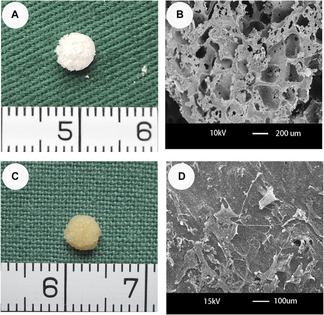
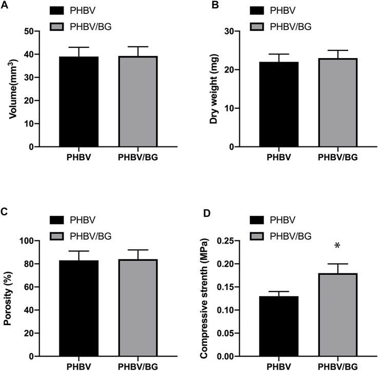
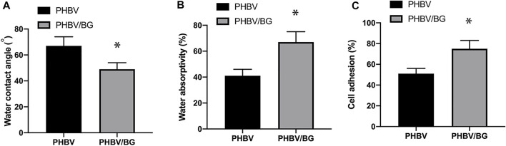
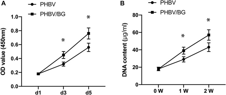
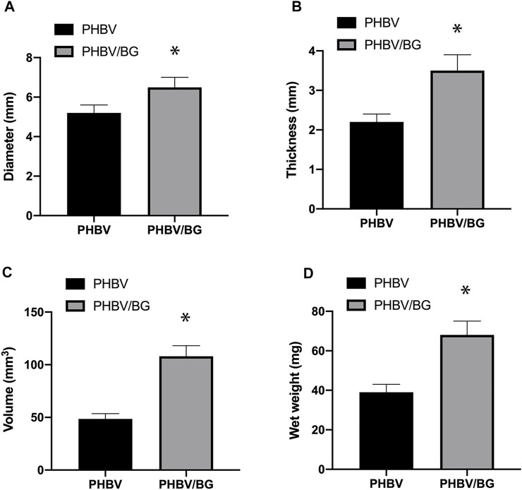
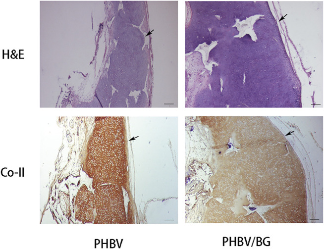
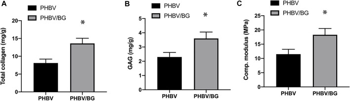
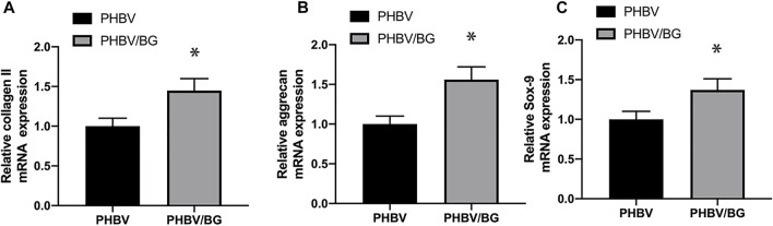
Background: The Poly (3-hydroxybutyrate-co-3-hydroxyvalerate) (PHBV) scaffold has proven to be a promising three-dimensional (3D) biodegradable and bioactive scaffold for the growth and proliferation of cartilage progenitor cells (CPCs). The addition of Bioglass into PHBV was reported to increase the bioactivity and mechanical properties of the bioactive materials. Methods: In the current study, the influence of the addition of Bioglass into PHBV 3D porous scaffolds on the characteristics of CPC-based tissue-engineered cartilages in vivo were compared. CPCs were seeded into 3D macroporous PHBV scaffolds and PHBV/10% Bioglass scaffolds. The CPC-scaffold constructs underwent 6 weeks in vitro chondrogenic induction culture and were then transplanted in vivo for another 6 weeks to evaluate the difference between the CPC-PHBV construct and CPC-PHBV/10% Bioglass construct in vivo. Results: Compared with the pure PHBV scaffold, the PHBV/10% Bioglass scaffold has better hydrophilicity and a higher percentage of adhered cells. The CPC-PHBV/10%Bioglass construct produced much more cartilage-like tissues with higher cartilage-relative gene expression and cartilage matrix protein production and better biomechanical performance than the CPC-PHBV construct. Conclusion: The addition of Bioglass into 3D PHBV macroporous scaffolds improves the characteristics of CPC-based tissue-engineered cartilages in vivo.
Keywords: Bioglass; PHBV; cartilage engineering; cartilage progenitor cells; hydrophilicity.
Copyright © 2022 Xue, Zhang, Ge, Wang, Qi and Liu.
Conflict of interest statement
The authors declare that the research was conducted in the absence of any commercial or financial relationships that could be construed as a potential conflict of interest.
Figures
Similar articles
-
Biomedical Applications of the Biopolymer Poly(3-hydroxybutyrate-co-3-hydroxyvalerate) (PHBV): Drug Encapsulation and Scaffold Fabrication.Int J Mol Sci. 2023 Jul 19;24(14):11674. doi: 10.3390/ijms241411674. Int J Mol Sci. 2023. PMID: 37511432 Free PMC article. Review.
-
Improvement of PHBV scaffolds with bioglass for cartilage tissue engineering.PLoS One. 2013 Aug 9;8(8):e71563. doi: 10.1371/journal.pone.0071563. eCollection 2013. PLoS One. 2013. PMID: 23951190 Free PMC article.
-
Cartilage progenitor cells combined with PHBV in cartilage tissue engineering.J Transl Med. 2019 Mar 29;17(1):104. doi: 10.1186/s12967-019-1855-x. J Transl Med. 2019. PMID: 30925884 Free PMC article.
-
Cartilage tissue engineering using PHBV and PHBV/Bioglass scaffolds.Mol Med Rep. 2014 Jul;10(1):508-14. doi: 10.3892/mmr.2014.2145. Epub 2014 Apr 15. Mol Med Rep. 2014. PMID: 24737242
-
Design and fabrication of porous biodegradable scaffolds: a strategy for tissue engineering.J Biomater Sci Polym Ed. 2017 Nov;28(16):1797-1825. doi: 10.1080/09205063.2017.1354674. Epub 2017 Jul 24. J Biomater Sci Polym Ed. 2017. PMID: 28707508 Review.
Cited by
-
Achieving Nasal Septal Cartilage In Situ Regeneration: Focus on Cartilage Progenitor Cells.Biomolecules. 2023 Aug 25;13(9):1302. doi: 10.3390/biom13091302. Biomolecules. 2023. PMID: 37759702 Free PMC article. Review.
-
Injectable mesoporous bioactive glass/sodium alginate hydrogel loaded with melatonin for intervertebral disc regeneration.Mater Today Bio. 2023 Jul 17;22:100731. doi: 10.1016/j.mtbio.2023.100731. eCollection 2023 Oct. Mater Today Bio. 2023. PMID: 37533731 Free PMC article.
-
Biomedical Applications of the Biopolymer Poly(3-hydroxybutyrate-co-3-hydroxyvalerate) (PHBV): Drug Encapsulation and Scaffold Fabrication.Int J Mol Sci. 2023 Jul 19;24(14):11674. doi: 10.3390/ijms241411674. Int J Mol Sci. 2023. PMID: 37511432 Free PMC article. Review.
-
The application of bioglass to treat osteoarthritis.EXCLI J. 2023 Dec 4;22:1232-1234. doi: 10.17179/excli2023-6613. eCollection 2023. EXCLI J. 2023. PMID: 38234972 Free PMC article. No abstract available.
References
LinkOut - more resources
Full Text Sources