

Abdominal wall endometriosis: a case report
- PMID: 35685105
- PMCID: PMC9146603
- DOI: 10.11604/pamj.2022.41.193.33536
Abdominal wall endometriosis: a case report
Abstract
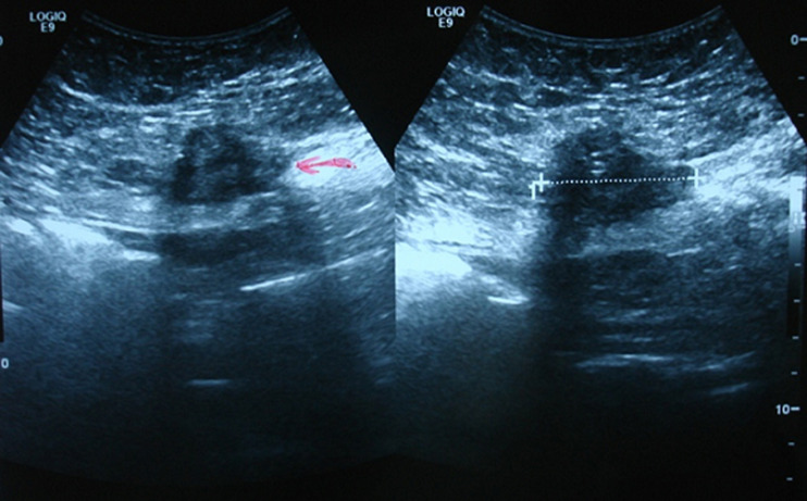
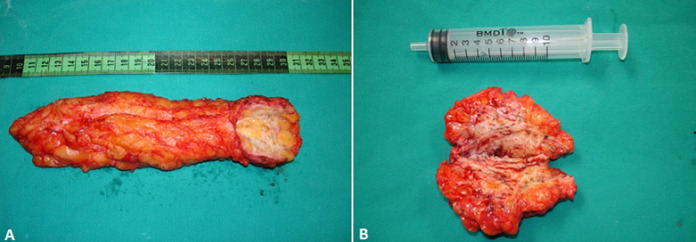
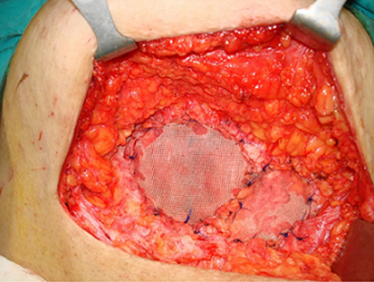
Abdominal wall endometriosis is the development of endometrial tissue in the anterior abdomen usually due to an operation in which the uterus is manipulated. We herein delineate the presentation, clinical investigation, and surgical treatment of an abdominal wall endometriosis case. A 42-year-old female presented with acute abdominal pain in the lower quadrants in the margins of an old cesarean scar. Two masses in the abdominal wall highly suspected of consisting of endometrial tissue were found during the investigation of the patient. These ones were removed in surgery and endometrial tissue secondary to previous cesarean section was confirmed after histological analysis. Consequently, although rare, if a painful mass in a surgical scar, such as a Pfannenstiel incision, is found in women of reproductive age with a history of obstetric surgery, the differential diagnosis shall include endometriosis. There is a portion of cases in which endometriosis recurs within five years following conservative surgery.
Keywords: Endometriosis; abdominal wall mass; case report; endometrioma; rectus abdominus.
Copyright: Daniel Paramythiotis et al.
Conflict of interest statement
The authors declare no competing interests.
Figures

Similar articles
-
Abdominal Wall Endometriosis at the Cesarean Section Scar.Pril (Makedon Akad Nauk Umet Odd Med Nauki). 2023 Dec 18;44(3):121-126. doi: 10.2478/prilozi-2023-0055. Print 2023 Dec 1. Pril (Makedon Akad Nauk Umet Odd Med Nauki). 2023. PMID: 38109456
-
Abdominal Wall Endometriosis Eleven Years After Cesarean Section: Case Report.Acta Clin Croat. 2017 Mar;56(1):162-165. doi: 10.20471/acc.2017.56.01.22. Acta Clin Croat. 2017. PMID: 29120557
-
Abdominal wall endometriosis following cesarean section: report of five cases.Clin Exp Obstet Gynecol. 2011;38(3):288-90. Clin Exp Obstet Gynecol. 2011. PMID: 21995169
-
Scar Endometriosis: a Case Report with Literature Review.Acta Med Iran. 2015 Dec;53(12):793-5. Acta Med Iran. 2015. PMID: 26749239 Review.
-
Abdominal scar endometriosis: A case report and review of literature.Indian J Pathol Microbiol. 2023 Oct-Dec;66(4):871-873. doi: 10.4103/ijpm.ijpm_460_22. Indian J Pathol Microbiol. 2023. PMID: 38084553 Review.
Cited by
-
Abdominal wall endometrioma after Cesarean section: a case series.J Surg Case Rep. 2024 May 7;2024(5):rjae295. doi: 10.1093/jscr/rjae295. eCollection 2024 May. J Surg Case Rep. 2024. PMID: 38721261 Free PMC article.
References
-
- Zondervan KT, Becker CM, Koga K, Missmer SA, Taylor RN, Viganò P. Endometriosis. Nat Rev Dis Primers. 2018 Jul 19;4(1):9. - PubMed
-
- Falcone T, Flyckt R. Clinical management of endometriosis. Obstet Gynecol. 2018;131(3):557–571. - PubMed
-
- Guerriero S, Condous G, van den Bosch T, Valentin L, Leone FPG, Van Schoubroeck D, et al. Systematic approach to sonographic evaluation of the pelvis in women with suspected endometriosis, including terms, definitions and measurements: a consensus opinion from the International Deep Endometriosis Analysis (IDEA) group. Ultrasound Obstet Gynecol. 2016;48(3):318–332. - PubMed
Publication types
MeSH terms
LinkOut - more resources
Full Text Sources
Medical
