

Inner Ear Drug Delivery for Sensorineural Hearing Loss: Current Challenges and Opportunities
- PMID: 35685768
- PMCID: PMC9170894
- DOI: 10.3389/fnins.2022.867453
Inner Ear Drug Delivery for Sensorineural Hearing Loss: Current Challenges and Opportunities
Abstract
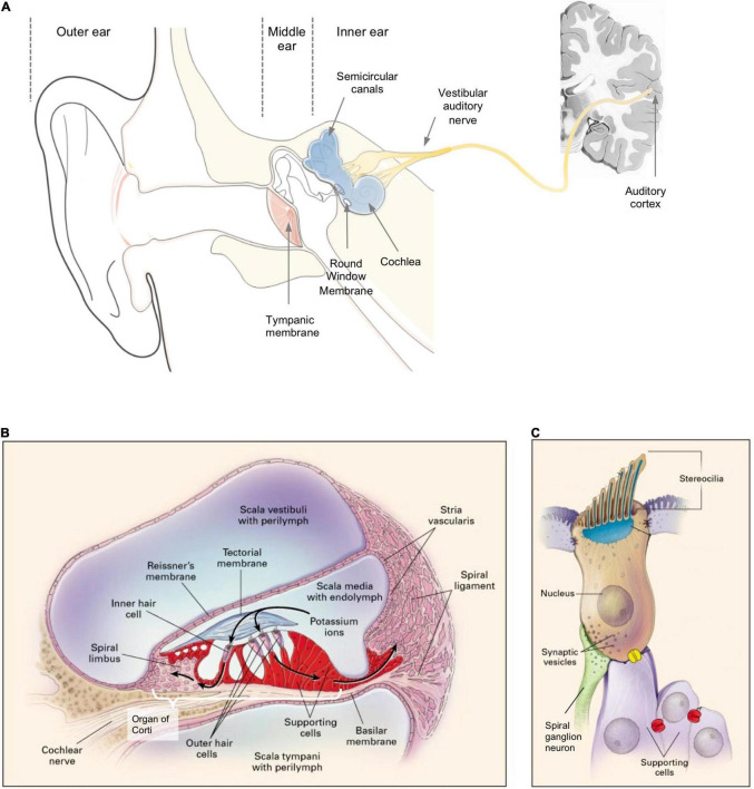
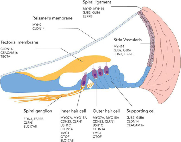
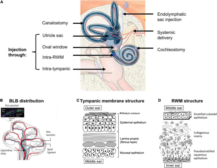
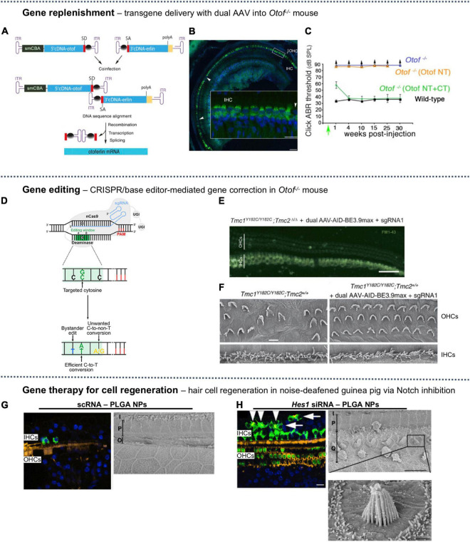
Most therapies for treating sensorineural hearing loss are challenged by the delivery across multiple tissue barriers to the hard-to-access anatomical location of the inner ear. In this review, we will provide a recent update on various pharmacotherapy, gene therapy, and cell therapy approaches used in clinical and preclinical studies for the treatment of sensorineural hearing loss and approaches taken to overcome the drug delivery barriers in the ear. Small-molecule drugs for pharmacotherapy can be delivered via systemic or local delivery, where the blood-labyrinth barrier hinders the former and tissue barriers including the tympanic membrane, the round window membrane, and/or the oval window hinder the latter. Meanwhile, gene and cell therapies often require targeted delivery to the cochlea, which is currently achieved via intra-cochlear or intra-labyrinthine injection. To improve the stability of the biomacromolecules during treatment, e.g., RNAs, DNAs, proteins, additional packing vehicles are often required. To address the diverse range of biological barriers involved in inner ear drug delivery, each class of therapy and the intended therapeutic cargoes will be discussed in this review, in the context of delivery routes commonly used, delivery vehicles if required (e.g., viral and non-viral nanocarriers), and other strategies to improve drug permeation and sustained release (e.g., hydrogel, nanocarriers, permeation enhancers, and microfluidic systems). Overall, this review aims to capture the important advancements and key steps in the development of inner ear therapies and delivery strategies over the past two decades for the treatment and prophylaxis of sensorineural hearing loss.
Keywords: cell therapy; drug delivery; gene therapy; inner ear; sensorineural hearing loss; small molecule.
Copyright © 2022 Liu and Yang.
Conflict of interest statement
The authors declare that the research was conducted in the absence of any commercial or financial relationships that could be construed as a potential conflict of interest.
Figures
Similar articles
-
Drug delivery to the inner ear: strategies and their therapeutic implications for sensorineural hearing loss.Curr Drug Deliv. 2012 May;9(3):231-42. doi: 10.2174/156720112800389098. Curr Drug Deliv. 2012. PMID: 22283653 Review.
-
Overcoming barriers: a review on innovations in drug delivery to the middle and inner ear.Front Pharmacol. 2023 Oct 19;14:1207141. doi: 10.3389/fphar.2023.1207141. eCollection 2023. Front Pharmacol. 2023. PMID: 37927600 Free PMC article. Review.
-
Nanodelivery of antioxidant Agents: A promising strategy for preventing sensorineural hearing loss.Eur J Pharm Biopharm. 2024 Sep;202:114393. doi: 10.1016/j.ejpb.2024.114393. Epub 2024 Jul 9. Eur J Pharm Biopharm. 2024. PMID: 38992481 Review.
-
Application of Nanomedicine in Inner Ear Diseases.Front Bioeng Biotechnol. 2022 Feb 11;9:809443. doi: 10.3389/fbioe.2021.809443. eCollection 2021. Front Bioeng Biotechnol. 2022. PMID: 35223817 Free PMC article. Review.
-
Rhesus Cochlear and Vestibular Functions Are Preserved After Inner Ear Injection of Saline Volume Sufficient for Gene Therapy Delivery.J Assoc Res Otolaryngol. 2017 Aug;18(4):601-617. doi: 10.1007/s10162-017-0628-6. Epub 2017 Jun 23. J Assoc Res Otolaryngol. 2017. PMID: 28646272 Free PMC article.
Cited by
-
Hearing loss during chemotherapy: prevalence, mechanisms, and protection.Am J Cancer Res. 2024 Sep 25;14(9):4597-4632. doi: 10.62347/OKGQ4382. eCollection 2024. Am J Cancer Res. 2024. PMID: 39417180 Free PMC article. Review.
-
Decreasing the physical gap in the neural-electrode interface and related concepts to improve cochlear implant performance.Front Neurosci. 2024 Jul 24;18:1425226. doi: 10.3389/fnins.2024.1425226. eCollection 2024. Front Neurosci. 2024. PMID: 39114486 Free PMC article. Review.
-
VDAC1 Inhibition Protects Against Noise-Induced Hearing Loss via the PINK1/Parkin Pathway.CNS Neurosci Ther. 2025 Apr;31(4):e70410. doi: 10.1111/cns.70410. CNS Neurosci Ther. 2025. PMID: 40285415 Free PMC article.
-
Fucoidan-based Nanomedicine for Hearing Loss: Emerging Roles as Carrier and Therapeutic Agent.Pharm Res. 2025 Aug 19. doi: 10.1007/s11095-025-03912-5. Online ahead of print. Pharm Res. 2025. PMID: 40830728 Review.
-
Magnetic Targeting of AAV Gene Therapy for Inner Ear Following Systemic Delivery: Preliminary Findings and Transduction Pattern in Rat Cochlea.J Assoc Res Otolaryngol. 2025 Sep 8. doi: 10.1007/s10162-025-01009-9. Online ahead of print. J Assoc Res Otolaryngol. 2025. PMID: 40921905
References
Publication types
LinkOut - more resources
Full Text Sources
Other Literature Sources
Miscellaneous