

vol2Brain: A New Online Pipeline for Whole Brain MRI Analysis
- PMID: 35685943
- PMCID: PMC9171328
- DOI: 10.3389/fninf.2022.862805
vol2Brain: A New Online Pipeline for Whole Brain MRI Analysis
Abstract
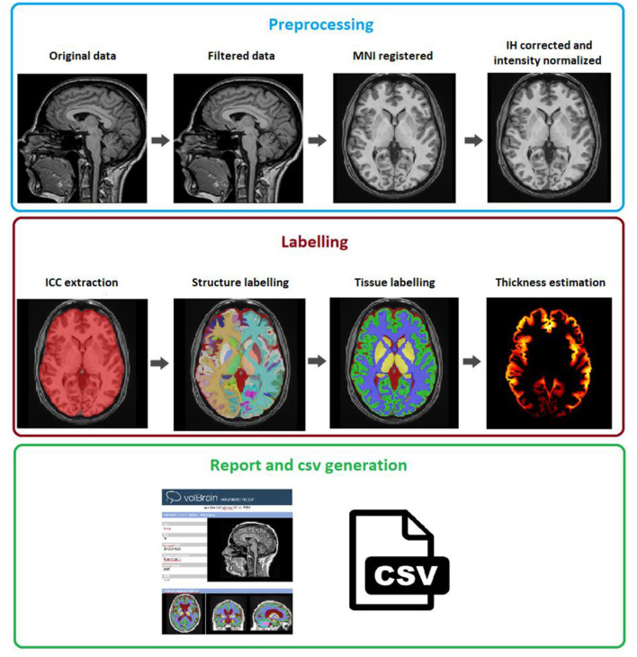
Automatic and reliable quantitative tools for MR brain image analysis are a very valuable resource for both clinical and research environments. In the past few years, this field has experienced many advances with successful techniques based on label fusion and more recently deep learning. However, few of them have been specifically designed to provide a dense anatomical labeling at the multiscale level and to deal with brain anatomical alterations such as white matter lesions (WML). In this work, we present a fully automatic pipeline (vol2Brain) for whole brain segmentation and analysis, which densely labels (N > 100) the brain while being robust to the presence of WML. This new pipeline is an evolution of our previous volBrain pipeline that extends significantly the number of regions that can be analyzed. Our proposed method is based on a fast and multiscale multi-atlas label fusion technology with systematic error correction able to provide accurate volumetric information in a few minutes. We have deployed our new pipeline within our platform volBrain (www.volbrain.upv.es), which has been already demonstrated to be an efficient and effective way to share our technology with the users worldwide.
Keywords: MRI; analysis; brain; cloud; segmentation.
Copyright © 2022 Manjón, Romero, Vivo-Hernando, Rubio, Aparici, de la Iglesia-Vaya and Coupé.
Conflict of interest statement
The authors declare that the research was conducted in the absence of any commercial or financial relationships that could be construed as a potential conflict of interest.
Figures
Similar articles
-
volBrain: An Online MRI Brain Volumetry System.Front Neuroinform. 2016 Jul 27;10:30. doi: 10.3389/fninf.2016.00030. eCollection 2016. Front Neuroinform. 2016. PMID: 27512372 Free PMC article.
-
A computational pipeline for quantification of pulmonary infections in small animal models using serial PET-CT imaging.EJNMMI Res. 2013 Jul 23;3(1):55. doi: 10.1186/2191-219X-3-55. EJNMMI Res. 2013. PMID: 23879987 Free PMC article.
-
An anatomical knowledge-based MRI deep learning pipeline for white matter hyperintensity quantification associated with cognitive impairment.Comput Med Imaging Graph. 2021 Apr;89:101873. doi: 10.1016/j.compmedimag.2021.101873. Epub 2021 Feb 3. Comput Med Imaging Graph. 2021. PMID: 33610084
-
Hierarchical multi-atlas label fusion with multi-scale feature representation and label-specific patch partition.Neuroimage. 2015 Feb 1;106:34-46. doi: 10.1016/j.neuroimage.2014.11.025. Epub 2014 Nov 20. Neuroimage. 2015. PMID: 25463474 Free PMC article.
-
Multiseg pipeline: automatic tissue segmentation of brain MR images with subject-specific atlases.Proc SPIE Int Soc Opt Eng. 2019 Feb;10953:109530K. doi: 10.1117/12.2513237. Epub 2019 Mar 15. Proc SPIE Int Soc Opt Eng. 2019. PMID: 31057202 Free PMC article.
Cited by
-
Changes in the volumes and asymmetry of subcortical structures in healthy individuals according to gender.Anat Sci Int. 2023 Sep;98(4):506-519. doi: 10.1007/s12565-023-00714-w. Epub 2023 Mar 22. Anat Sci Int. 2023. PMID: 36947348
-
Measurement of Limbic System Anatomical Volumes in Patients Diagnosed with Schizophrenia Using Vol2brain and Comparison with Healthy Individuals.Medicina (Kaunas). 2025 Mar 17;61(3):525. doi: 10.3390/medicina61030525. Medicina (Kaunas). 2025. PMID: 40142336 Free PMC article.
-
Reversal of cerebral pseudoatrophy in normal pressure hydrocephalus after ventriculoatrial shunt placement.Sci Rep. 2025 Jul 10;15(1):24817. doi: 10.1038/s41598-025-03158-6. Sci Rep. 2025. PMID: 40640209 Free PMC article.
-
Scan-rescan reliability assessment of brain volumetric analysis across scanners and software solutions.Sci Rep. 2025 Aug 14;15(1):29843. doi: 10.1038/s41598-025-15283-3. Sci Rep. 2025. PMID: 40813790 Free PMC article.
-
Heritability of Subcortical Grey Matter Structures.Medicina (Kaunas). 2022 Nov 21;58(11):1687. doi: 10.3390/medicina58111687. Medicina (Kaunas). 2022. PMID: 36422226 Free PMC article.
References
LinkOut - more resources
Full Text Sources
Miscellaneous