. 2022 May 24:12:887210.
doi: 10.3389/fonc.2022.887210. eCollection 2022.

Extracellular Vesicles: A New Source of Biomarkers in Pediatric Solid Tumors? A Systematic Review

Affiliations

Extracellular Vesicles: A New Source of Biomarkers in Pediatric Solid Tumors? A Systematic Review

Nathalie S M Lak et al. Front Oncol. .

Abstract

Virtually every cell in the body releases extracellular vesicles (EVs), the contents of which can provide a "fingerprint" of their cellular origin. EVs are present in all bodily fluids and can be obtained using minimally invasive techniques. Thus, EVs can provide a promising source of diagnostic, prognostic, and predictive biomarkers, particularly in the context of cancer. Despite advances using EVs as biomarkers in adult cancers, little is known regarding their use in pediatric cancers. In this review, we provide an overview of published clinical and in vitro studies in order to assess the potential of using EV-derived biomarkers in pediatric solid tumors. We performed a systematic literature search, which yielded studies regarding desmoplastic small round cell tumor, hepatoblastoma, neuroblastoma, osteosarcoma, and rhabdomyosarcoma. We then determined the extent to which the in vivo findings are supported by in vitro data, and vice versa. We also critically evaluated the clinical studies using the GRADE (Grading of Recommendations Assessment, Development, and Evaluation) system, and we evaluated the purification and characterization of EVs in both the in vivo and in vitro studies in accordance with MISEV guidelines, yielding EV-TRACK and PedEV scores. We found that several studies identified similar miRNAs in overlapping and distinct tumor entities, indicating the potential for EV-derived biomarkers. However, most studies regarding EV-based biomarkers in pediatric solid tumors lack a standardized system of reporting their EV purification and characterization methods, as well as validation in an independent cohort, which are needed in order to bring EV-based biomarkers to the clinic.

Keywords: desmoplastic small round cell tumor; extracellular vesicles; hepatoblastoma; neuroblastoma; osteosarcoma; pediatric oncology; rhabdomyosarcoma; solid tumors.


Conflict of interest statement

The authors declare that the research was conducted in the absence of any commercial or financial relationships that could be construed as a potential conflict of interest.

Figures

Figure 1
Figure 1
Extracellular vesicles (EVs) from blood as a liquid biopsy: isolation methods and downstream analyses. Left: EVs (including tumor-derived EVs) are isolated from peripheral blood and purified using differential centrifugation/ultracentrifugation (A), size exclusion chromatography [SEC; (B)], density gradient (C), commercially available precipitating agents [e.g., Exoquick; (D)], immunoprecipitation/capture (E) or microfluidic/nanostructure approaches (F). Right, top panel: the isolated EVs are then characterized using (from the top-left, moving clockwise) electron microscopy, flow cytometry, western blot analysis, and/or nanoparticle tracking analysis (NTA). Right, bottom panel: the EV contents are analyzed using (from the top left, moving clockwise) mass spectrometry, RNA sequencing, enzyme-linked immunosorbent assay (ELISA), and/or RT-qPCR.
Figure 2
Figure 2
Number of papers published in the indicated years regarding extracellular vesicles (EVs), pediatric solid tumors, tumor-derived EVs, and both EVs and pediatric solid tumors. The inset shows only the publications regarding both EVs and pediatric solid tumors.
Figure 3
Figure 3
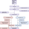
Flow diagram depicting the search strategy and inclusion and exclusion of studies. JEV, Journal of Extracellular Vesicles.
Figure 4
Figure 4
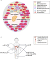
(A) Overview of the hallmarks of cancer and the differentially regulated miRNAs described in the various in vitro and in vivo reports, classified according to their function. DSRCT, desmoplastic small round cell tumor (B) Differentially regulated miRNAs in the indicated solid tumors (hepatoblastoma, neuroblastoma, DSRCT, and osteosarcoma) based on the in vivo and in vitro publications (↑, upregulated; ↓, downregulated). References for miR-21 (65, 79, 80, 85, 86); for miR-25-3p (80, 82, 83); for miR199a-3p (67, 69):; for miR-34 (64, 66); for miR92a (73, 80) and for miR-342-3p (64, 68).

References

    1. van Niel G, D’Angelo G, Raposo G. Shedding Light on the Cell Biology of Extracellular Vesicles. Nat Rev Mol Cell Biol (2018) 19(4):213–28. doi: 10.1038/nrm.2017.125 - DOI - PubMed
    1. Bebelman MP, Smit MJ, Pegtel DM, Baglio SR. Biogenesis and Function of Extracellular Vesicles in Cancer. Pharmacol Ther (2018) 188:1–11. doi: 10.1016/j.pharmthera.2018.02.013 - DOI - PubMed
    1. Becker A, Thakur BK, Weiss JM, Kim HS, Peinado H, Lyden D. Extracellular Vesicles in Cancer: Cell-To-Cell Mediators of Metastasis. Cancer Cell (2016) 30(6):836–48. doi: 10.1016/j.ccell.2016.10.009 - DOI - PMC - PubMed
    1. van Eijndhoven MA, Zijlstra JM, Groenewegen NJ, Drees EE, van Niele S, Baglio SR, et al. . Plasma Vesicle miRNAs for Therapy Response Monitoring in Hodgkin Lymphoma Patients. JCI Insight (2016) 1(19):e89631. doi: 10.1172/jci.insight.89631 - DOI - PMC - PubMed
    1. Merchant ML, Rood IM, Deegens JKJ, Klein JB. Isolation and Characterization of Urinary Extracellular Vesicles: Implications for Biomarker Discovery. Nat Rev Nephrol (2017) 13(12):731–49. doi: 10.1038/nrneph.2017.148 - DOI - PMC - PubMed

Publication types

LinkOut - more resources