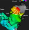
Detection of the origin of atrial tachycardia by 3D electro-anatomical mapping and treatment by radiofrequency catheter ablation in horses
- PMID: 35686355
- PMCID: PMC9308432
- DOI: 10.1111/jvim.16473
Detection of the origin of atrial tachycardia by 3D electro-anatomical mapping and treatment by radiofrequency catheter ablation in horses
Abstract
Background: Atrial tachycardia (AT) can be treated by medical or electrical cardioversion but the recurrence rate is high. Three-dimensional electro-anatomical mapping, recently described in horses, might be used to map AT to identify a focal source or reentry mechanism and to guide treatment by radiofrequency ablation.
Objectives: To describe the feasibility of 3D electro-anatomical mapping and radiofrequency catheter ablation to characterize and treat sustained AT in horses.
Animals: Nine horses with sustained AT.
Methods: Records from horses with sustained AT referred for radiofrequency ablation at Ghent University were reviewed.
Results: The AT was drug resistant in 4 out of 9 horses. In 8 out of 9 horses, AT originated from a localized macro-reentrant circuit (n = 5) or a focal source (n = 3) located at the transition between the right atrium and the caudal vena cava. In these 8 horses, local radiofrequency catheter ablation resulted in the termination of AT. At follow-up, 6 out of 8 horses remained free of recurrence.
Conclusions and clinical importance: Differentiation between focal and macro-reentrant AT in horses is possible using 3D electro-anatomical mapping. In this study, the source of right atrial AT in horses was safely treated by radiofrequency catheter ablation.
Keywords: arrhythmia; atrial flutter; electrophysiology; focal atrial tachycardia; supraventricular tachyarrhythmia.
© 2022 The Authors. Journal of Veterinary Internal Medicine published by Wiley Periodicals LLC on behalf of American College of Veterinary Internal Medicine.
Conflict of interest statement
Authors declare no conflict of interest.
Figures


References
-
- van Loon G. Cardiac arrhythmias in horses. Vet Clin N Am Equine Pract. 2019;35(1):85‐102. - PubMed
-
- van Steenkiste G, de Clercq D, Vera L, Decloedt A, van Loon G. Sustained atrial tachycardia in horses and treatment by transvenous electrical cardioversion. Equine Vet J. 2019;51:634‐640. - PubMed
-
- Whelchel DD, Tennent‐Brown BS, Coleman AE, et al. Treatment of supraventricular tachycardia in a horse. J Vet Emerg Crit Care. 2017;27(3):362‐368. - PubMed
-
- van Loon G, Jordaens L, Muylle E, Nollet G, Sustronck B. Intracardiac overdrive pacing as a treatment of atrial flutter in a horse. Vet Rec. 1998;142:301‐303. - PubMed
-
- Saoudi N, Cosio F, Waldo A, et al. Classification of atrial flutter and regular atrial tachycardia according to electrophysiologic mechanism and anatomic bases: a statement from a Joint Expert Group from the Working Group of Arrhythmias of the European Society of Cardiology and the North American Society of Pacing and Electrophysiology. J Cardiovasc Electrophysiol. 2001;22(14):1162‐1182. - PubMed
MeSH terms
Grants and funding
LinkOut - more resources
Full Text Sources

