. 2022 Sep;67(3):334-345.
doi: 10.1165/rcmb.2021-0548OC.

Secretory Cells Are the Primary Source of pIgR in Small Airways

Affiliations

Secretory Cells Are the Primary Source of pIgR in Small Airways

Jessica B Blackburn et al. Am J Respir Cell Mol Biol. 2022 Sep.

Abstract

Loss of secretory IgA (SIgA) is common in chronic obstructive pulmonary disease (COPD) small airways and likely contributes to disease progression. We hypothesized that loss of SIgA results from reduced expression of pIgR (polymeric immunoglobulin receptor), a chaperone protein needed for SIgA transcytosis, in the COPD small airway epithelium. pIgR-expressing cells were defined and quantified at single-cell resolution in human airways using RNA in situ hybridization, immunostaining, and single-cell RNA sequencing. Complementary studies in mice used immunostaining, primary murine tracheal epithelial cell culture, and transgenic mice with secretory or ciliated cell-specific knockout of pIgR. SIgA degradation by human neutrophil elastase or secreted bacterial proteases from nontypeable Haemophilus influenzae was evaluated in vitro. We found that secretory cells are the predominant cell type responsible for pIgR expression in human and murine airways. Loss of SIgA in small airways was not associated with a reduction in secretory cells but rather a reduction in pIgR protein expression despite intact PIGR mRNA expression. Neutrophil elastase and nontypeable H. influenzae-secreted proteases are both capable of degrading SIgA in vitro and may also contribute to a deficient SIgA immunobarrier in COPD. Loss of the SIgA immunobarrier in small airways of patients with severe COPD is complex and likely results from both pIgR-dependent defects in IgA transcytosis and SIgA degradation.

Keywords: airway epithelium; chronic obstructive pulmonary disease; secretory IgA; secretory cells.


Figures

Figure 1.
Figure 1.
PIGR/pIgR (polymeric immunoglobulin receptor) is expressed primarily by secretory cells in human lung sections. (A) RNA in situ hybridization for PIGR (red), the secretory cell marker SCGB1A1 (secretoglobin family 1A member 1) (green, left panel), the ciliated cell marker FOXJ1 (forkhead box J1) (green, right panel), and DAPI (white) in small airways from deceased lung donors without chronic respiratory disease. Scale bars, 50 μm. Insets depict 2.5× magnification. (B) FOXJ1, SCGB1A1, and PIGR expression (probe counts) in cells defined as SCGB1A1+ and FOXJ1+; anything less than 10 probes per cell was considered background or overlap from adjacent cells. Error bars represent median values and 95% confidence interval. *P < 0.0001 (Mann-Whitney test). (C) RNA in situ hybridization showing heterogeneous expression of PIGR (blue), SCGB1A1 (green), and SCGB3A2 (secretoglobin family 3A member 2) (red) and the combination of each marker in a small airway from a control patient. Scale bar, 50 μm. (D) Percentage of cells expressing PIGR alone or PIGR with secretory cell markers SCGB1A1 and/or SCGB3A2; n = 10,908 cells (4,571 PIGR expressing) from 17 airways in six deceased lung donors without chronic respiratory disease. (E) Immunostaining for pIgR (red), Scgb1a1 (green, left panel), FoxJ1 (green, center panel), and all three markers (pIgR, red; Scgb1a1, green; FoxJ1, blue; DAPI, white; right panel) in small airways from deceased lung donors without chronic respiratory disease. Scale bars, 50 μm. Insets depict 2× magnification.
Figure 2.
Figure 2.
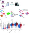
PIGR is expressed primarily by secretory cells in human lung tissue by single-cell RNA sequencing (scRNA-seq). (A) Tissue processing work flow for scRNA-seq experiments. (B) Uniform manifold approximation and projection (UMAP)–embedded image showing cell clusters derived from nonbiased, graph-based clustering of EPCAM+ (epithelial cell adhesion molecule) cells derived from the distal lungs of patients with chronic obstructive pulmonary disease (COPD) and control patients. (C) UMAP-embedded image showing integration of cells according to clinical status. (D) Violin plots show PIGR expression in COPD explants and organ donors without known chronic respiratory disease (e.g., control subjects). The height of each violin corresponds to the degree of expression/cell, and the width of the violin corresponds to the number of expressing cells for a given amount of expression. *P < 0.001 and **P < 0.0001 (negative binomial regression). AT1 = alveolar type 1; AT2 = alveolar type 2; KRT5 = keratin 5; KRT17 = keratin 17; TGen = Translational Genomics Research Institute; VUMC = Vanderbilt University Medical Center.
Figure 3.
Figure 3.
pIgR is expressed primarily by secretory cells in murine airways. (A) Immunostaining for pIgR (red), ciliated cell marker acetylated-α tubulin (white), and secretory cell marker Scgb1a1 (green) in airways from wild-type C57BL6/J mice. DAPI staining of nuclei (DNA) is shown in blue. Scale bars, 20 μm. (B) Diagram describing methods used for DAPT treatment in primary murine tracheal epithelial cells (MTECs). (C) IB for pIgR in air–liquid interface–differentiated MTECs with and without addition of the γ-secretase inhibitor DAPT, which restricts secretory cell differentiation (n = 3 inserts per group). β-Actin is included as a loading control, and α-tubulin is included to show that DAPT did not affect ciliated cell differentiation. (D) Quantification of (C) by densitometry. All three bands of pIgR are included in the densitometry analysis. pIgR expression was normalized across samples using β-actin. *P < 0.01 (t test). SC = secretory component; WT = wild-type.
Figure 4.
Figure 4.
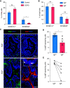
Ciliated cell–derived pIgR is dispensable for protection against emphysema. (A and B) Immunostaining for pIgR and GFP in adult pIgRΔciliated and pIgRΔsecretory mice. White arrows in (A) highlight pIgR+GFP cells in pIgRΔciliated mice. Scale bars, 50 μm. Inset depicts 2× magnification. (C) Low-magnification images (10×) of the lung parenchyma in 6-month-old Cre and Cre+ pIgRΔciliated mice shows absence of emphysema in both strains (hematoxylin and eosin stain). (D) Quantification of emphysema (by mean linear intercept) in 6-month-old C57Bl6 WT and Cre+ and Cre pIgRΔciliated mice (n = 4–7 mice per group). Box-and-whisker plots represent median, interquartile range, and range. Results are not significant (ANOVA). ns = not significant.
Figure 5.
Figure 5.
Secretory IgA (SIgA) airways have reduced pIgR expression compared with SIgA+ airways in patients with advanced COPD. (A) Percentage of SCGB1A1+ or SCGB3A2+ cells per airway among DAPI+ cells as assessed using RNA-ISH in control lungs (n = 6; 17 airways) versus COPD (n = 6; 18 airways). *P < 0.05, Mann-Whitney test. (B) Percentage of SCGB1A1+ or SCGB3A2+ cells per airway among DAPI+ cells as assessed using RNA-ISH in SIgA+ and SIgA airways (five patients, 18 positive airways, 28 negative airways) as shown in (C). (C) Representative images of a SIgA+ and SIgA small airways (on the basis of IgA immunostaining) from the explanted lungs of a patient with COPD (left) and staining of pIgR in a serial section of the same airways showing reduced pIgR in SIgA-deficient airways (right). Scale bars, 50 μm. Insets depict 2× magnification. Arrows show that intracellular pIgR is still present. (D) Average percentage of pIgR-expressing cells per patient in SIgA+ or SIgA airways as determined on HALO analysis. *P < 0.05, Mann-Whitney test. (E) Paired analysis of percentage of pIgR-expressing cells in SIgA versus SIgA+ airways per patient. ns = not significant (P > 0.05); RNA-ISH = RNA in situ hybridization.
Figure 6.
Figure 6.
Human SIgA is degraded by bacterial and host proteases in vitro. (A) pIgR-positive, SIgA-negative airways were assessed for the presence of bacteria using 16S ribosomal targeted fluorescence in situ hybridization. Scale bars, 20 μm. (B) SIgA was incubated with conditioned media from NTHi strain 1479 for 24 hours and then assessed for degradation/cleavage products in the IgA heavy chain (top) or in the secretory component (bottom). PBS and noninoculated media were used as negative controls. (C) Quantification of full-length IgA heavy chain from the top panel of (B). (D) Quantification of full-length secretory component from the bottom panel of (B). (E) pIgR-positive, SIgA-negative airways were assessed for the presence of neutrophils as detected by the neutrophil protease elastase. Arrow in the center panel indicates that intracellular pIgR is still present. Inset depicts 2× magnification. Neutrophils are present both in (arrow) and surrounding (arrowhead) the airway. Scale bars, 100 μm. (F) SIgA was incubated with human neutrophil elastase (HNE) for 24 hours and then assessed for degradation/cleavage products in the IgA heavy chain (top) or in the secretory component (bottom). The buffer used to dissolve the HNE (vehicle) was used as a negative control. A nonspecific mammalian protease inhibitor was also incubated with SIgA and HNE as a control. (G) Quantification of full-length IgA heavy chain from the top panel of (F). (H) Quantification of full-length secretory component from the bottom panel of (F). *P < 0.01 (t test). NE = neutrophil elastase; NTHi = nontypeable Haemophilus influenzae; PI = protease inhibitor.

Comment in

References

    1. Combs MP, Dickson RP. Turning the lungs inside out: the intersecting microbiomes of the lungs and the built environment. Am J Respir Crit Care Med . 2020;202:1618–1620. - PMC - PubMed
    1. Muir DC. Bulk flow and diffusion in the airways of the lung. Br J Dis Chest . 1966;60:169–176. - PubMed
    1. Diamond G, Legarda D, Ryan LK. The innate immune response of the respiratory epithelium. Immunol Rev . 2000;173:27–38. - PubMed
    1. Ganesan S, Comstock AT, Sajjan US. Barrier function of airway tract epithelium. Tissue Barriers . 2013;1:e24997. - PMC - PubMed
    1. Kaetzel CS. The polymeric immunoglobulin receptor: bridging innate and adaptive immune responses at mucosal surfaces. Immunol Rev . 2005;206:83–99. - PubMed

Publication types

MeSH terms

Substances