. 2022 Aug 15;206(4):459-475.
doi: 10.1164/rccm.202109-2151OC.

CD38 Mediates Lung Fibrosis by Promoting Alveolar Epithelial Cell Aging

Affiliations

CD38 Mediates Lung Fibrosis by Promoting Alveolar Epithelial Cell Aging

Huachun Cui et al. Am J Respir Crit Care Med. .

Abstract

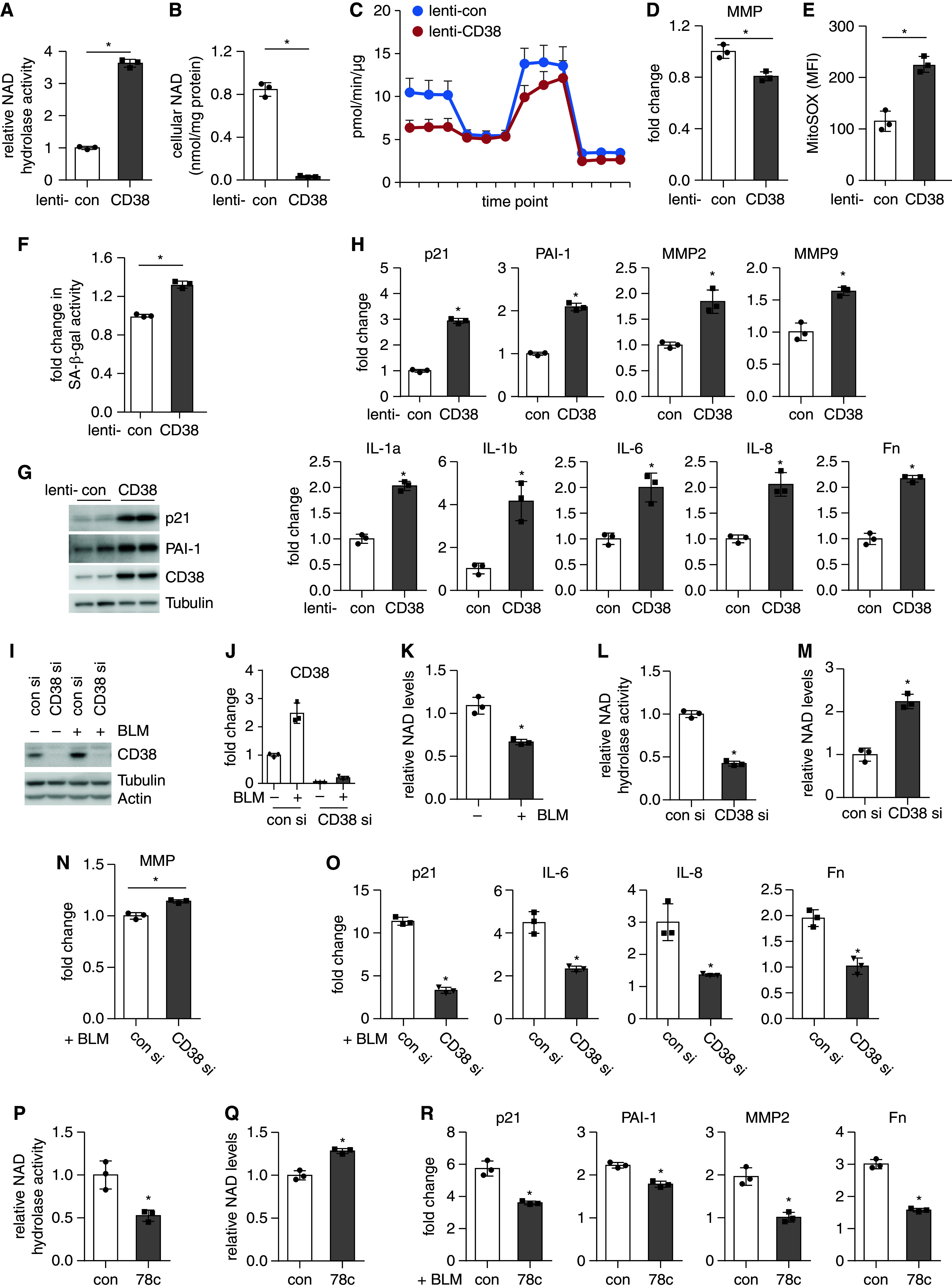
Rationale: A prevailing paradigm recognizes idiopathic pulmonary fibrosis (IPF) originating from various alveolar epithelial cell (AEC) injuries, and there is a growing appreciation of AEC aging as a key driver of the pathogenesis. Despite this progress, it is incompletely understood what main factor(s) contribute to the worsened alveolar epithelial aging in lung fibrosis. It remains a challenge how to dampen AEC aging and thereby mitigate the disease progression. Objectives: To determine the role of AEC CD38 (cluster of differentiation 38) in promoting cellular aging and lung fibrosis. Methods: We used single-cell RNA sequencing, real-time PCR, flow cytometry, and Western blotting. Measurements and Main Results: We discovered a pivotal role of CD38, a cardinal nicotinamide adenine dinucleotide (NAD) hydrolase, in AEC aging and its promotion of lung fibrosis. We found increased CD38 expression in IPF lungs that inversely correlated with the lung functions of patients. CD38 was primarily located in the AECs of human lung parenchyma and was markedly induced in IPF AECs. Similarly, CD38 expression was elevated in the AECs of fibrotic lungs of young mice and further augmented in those of old mice, which was in accordance with a worsened AEC aging phenotype and an aggravated lung fibrosis in the old animals. Mechanistically, we found that CD38 elevation downregulated intracellular NAD, which likely led to the aging promoting impairment of the NAD-dependent cellular and molecular activities. Furthermore, we demonstrated that genetic and pharmacological inactivation of CD38 improved these NAD dependent events and ameliorated bleomycin-induced lung fibrosis. Conclusions: Our study suggests targeting alveolar CD38 as a novel and effective therapeutic strategy to treat this pathology.

Keywords: CD38; alveolar epithelial cell; nicotinamide adenine dinucleotide; pulmonary fibrosis; senescence.


Figures

Figure 1.
Figure 1.
The elevated susceptibility to lung fibrosis with age is associated with a worsened alveolar epithelial senescence and mitochondrial dysfunction. (AC) Eight-week (young) and 18-month (old) male C57BL/6 mice were intratracheally instilled with saline or bleomycin (BLM, 1.5 U/kg in 50 μl saline). Three weeks after treatment, the concentrations of hydroxyproline (A), the extent of collagen deposition (B), and the concentrations of the indicated gene expression (C) in the lungs were determined; n = 3–6 mice for each group; mean ± SEM; *P < 0.05. Original magnification: ×20; scale bars: 100 μm for B. (DE) Young and old mice were treated as in AC. Three weeks after the treatment, primary alveolar epithelial cells (AECs) were isolated by fluorescence-activated cell sorting as described in Methods. Concentrations of indicated protein and genes in the AECs were determined by Western blotting (D) and real-time PCR (E). Densitometric analysis was performed with ImageJ (NIH) (D, bottom panel). n = 3–4 mice for each group; mean ± SEM; *P < 0.05. (FG) Young and old mice were treated as in AC. Three weeks after the treatment, primary AECs were isolated by magnetic activated cell sorting as described in Methods. 1 × 105 cells per well were plated in Matrigel precoated Seahorse XF-96 microplates. Real-time oxygen consumption rate was recorded and normalized by protein concentration. n = 4 mice for each group; mean ± SEM; *P < 0.05. (HJ) Young and old mice were treated and primary AECs isolated as in D and E. Cellular nicotinamide adenine dinucleotide (NAD) concentrations were measured. n = 3 mice for saline groups, n = 4–9 mice for bleomycin groups; mean ± SEM; *P < 0.05 and **P < 0.01. (K) Human normal control and idiopathic pulmonary fibrosis lung tissues were lysed. NAD concentrations in the lung lysates were measured and normalized by protein concentration. n = 10 for each group; mean ± SEM; *P < 0.05. Fn = fibronectin; IPF = idiopathic pulmonary fibrosis.
Figure 2.
Figure 2.
CD38, a cardinal nicotinamide adenine dinucleotide hydrolase, is primarily expressed in the alveolar epithelial cells (AECs) in the lung parenchyma and upregulated in the fibrotic mouse lungs. (AC) Eight-week (young) and 18-month (old) male C57BL/6 mice were intratracheally instilled with saline or bleomycin (BLM; 1.5 U/kg in 50 μl saline). Three weeks after treatment, mice were killed and primary AECs isolated from the lungs by fluorescence-activated cell sorting. AEC total RNAs were purified and the CD38 mRNA concentration assessed by real-time PCR A. AEC CD38 protein concentrations were determined by Western blotting, and densitometric analysis performed using ImageJ (B and C). n = 3–4 mice for each group; mean ± SEM; *P < 0.05. (D) Primary mouse AECs from wild-type mice and human lung epithelial A549 cells were treated with or without bleomycin (0.01 U/ml) for 2 days. Concentrations of the indicated proteins were determined by Western blotting. (EG) Young and old mice were treated with saline or bleomycin as in AC. Three weeks after treatment, mice were killed and lung sections prepared. Immunofluorescence staining and fluorescence microscopy were performed to determine the expression of CD38 and E-Cadherin. Nuclei were counterstained with DAPI. Original magnification: ×20; scale bars: 100 μm (EF). CD38 fluorescence intensity of the E-Cadherin+ AECs was quantified by ImageJ. Box-and-whisker plots: 25th and 75th percentiles, median, minimum, and maximum values. ***P < 0.001 (G). Con = control.
Figure 3.
Figure 3.
Alveolar epithelial cell CD38 is upregulated in idiopathic pulmonary fibrosis (IPF) lungs, and CD38 expression correlates inversely with the lung functions of patients with IPF. (A) Human CD38 mRNA expression in lung tissues from subjects with IPF (n = 160) and controls (n = 132) from the LGRC (Lung Genomics Research Consortium Cohort) was determined by microarray (GSE47460). (B) CD38 protein concentrations in normal controls and IPF lungs were determined by Western blotting and densitometric analysis performed by ImageJ. n = 7 for each group; mean ± SD; ***P < 0.001. (CD) Correlations of the lung CD38 expression with the FVC and Dl CO of the same cohort as in A are shown. (E) Uniform manifold approximation and projection (UMAP) of 8,942 resident lung parenchymal cells randomly downsampled from our previous dataset representative of 28 control and 32 IPF lung samples (GSE136831) (45). (F) UMAP with cells labeled by disease identity.(G) UMAP with cells labeled by normalized CD38 expression. (H) Boxplots representing the average normalized CD38 expression for each subject, grouped by cell type and disease status. Each dot represents one subject; whiskers are 1.5 × interquartile range. (I) Scatterplot of the average CD38 expression among alveolar type II (ATII) cells for each subject. A linear model is fitted to control subjects and patients with IPF; 95% SE confidence interval is projected over each line. Only subjects with at least 10 ATII cells sampled are included. (J and K) Sections of human normal control and IPF lungs were prepared. Immunofluorescence staining and fluorescence microscopy were performed to determine the expression and localization of CD38, surfactant protein C (SP-C), and smooth muscle actin-α (SMA-α). Nuclei were counterstained with DAPI (J). Immunohistochemistry staining for CD38 was performed in IPF lung slices (K). Original magnification: ×20 for J and ×10 for K. Scale bars: 100 μm for J and 200 μm for K. UIP = usual interstitial pneumonia.
Figure 4.
Figure 4.
CD38 promotes alveolar epithelial cell senescence and mitochondrial dysfunction in vitro. (A and B) Human lung epithelial A549 cells were transduced with control lentiviruses or viruses that express human CD38. Two to 3 days after infection, the cellular nicotinamide adenine dinucleotide (NAD) hydrolase activities and NAD concentrations were determined. n = 3 for each group; mean ± SD; *P < 0.05. (CE) Cells were treated as in A and B. Two to 3 days after infection, cells were subjected to mitochondrial oxygen consumption assay by XF-96 Bio-Analyzers (C), mitochondrial membrane potential (MMP) assay (D), and mitochondrial reactive oxygen species measurement (E). n = 3–4 for each group; mean ± SD; *P < 0.05. (FH) Cells were treated as in A and B. Two to 3 days after infection, senescence-associated β-galactosidase (SA-β-gal) activity (F) and concentrations of the indicated proteins (G) and genes (H) were determined by flow cytometry–based assay, Western blotting, or real-time PCR. n = 3 for each group; mean ± SD; *P < 0.05. (IO) Cells were transfected with control or human CD38 siRNAs. Two days after transfection, cells were treated with or without 0.01 U/ml bleomycin (BLM) for another 24–48 hours. The CD38 knockdown efficiency was determined by Western blotting and real-time PCR (I and J). Cellular NAD concentrations and NAD hydrolase activities were determined by the respective enzymatic assay (KM), MMP assessed by flow cytometry–based assay (N), and the expression of the indicated genes assessed by real-time PCR (O). n = 3 for each group; mean ± SD; *P < 0.05 for JO. (PR) Cells were pretreated with or without 78c (final concentration at 0.5 μM) for 2 hours, followed by bleomycin treatment (0.01 U/ml) for another 24–48 hours. Cells were harvested and cellular NAD hydrolase activities, NAD concentrations, and concentrations of the indicated genes were determined as in IO. n = 3 for each group; mean ± SD; *P < 0.05. Con = control; Fn = fibronectin; MFI = mean fluorescence intensity; si = siRNA.
Figure 5.
Figure 5.
CD38 regulates the activities of the nicotinamide adenine dinucleotide–dependent enzymes Sirt1 and Sirt3, key regulators of cellular aging, in alveolar epithelial cells (AECs). (AB) A549 human lung epithelial cells were transduced with control lentiviruses or viruses that express human CD38. Two to 3 days later, cellular Sirt1 and Sirt3 activities were examined. n = 3 for each group; mean ± SD; *P < 0.05. (C) Cells were treated as in A and B. Mitochondria were isolated, and mitochondria Sirt3 activities were assessed. n = 4 for each group; mean ± SD; *P < 0.05. (D) Cells were treated as in A and B. Total acetyl-lysine and concentrations of other indicated proteins were determined by Western blotting. (E and F) Cells were transfected with control or CD38 siRNAs. Two days later, cells were treated with or without 0.01 U/ml bleomycin (BLM) for another 24–48 hours. Cellular Sirt1 and Sirt3 activities were examined. n = 3 for each group; mean ± SD; *P < 0.05. (G and H) Cells were pretreated with or without 78c (0.5 μM) for 2 hours, followed by bleomycin treatment (0.01 U/ml) for another 48 hours. Cellular Sirt1 and Sirt3 activities were examined. n = 3 for each group; mean ± SD; *P < 0.05. (I) Cells were transfected with control or CD38 siRNAs. Two days later, cells were treated with 0.01 U/ml bleomycin for another 48 hours. Cellular total acetyl-lysine concentrations were determined by Western blotting. (J) Cells were pretreated with or without 78c for 2 hours, followed by 0.01 U/ml bleomycin treatment for another 48 hours. Cellular total acetyl-lysine concentrations were determined. (K and L) Primary AECs were isolated from young and old C57BL/6 mice by fluorescence-activated cell sorting. Cellular Sirt1 and Sirt3 activities were examined, and cellular total acetyl-lysine concentrations were determined by Western blotting. n = 3–4 mice for each group; mean ± SEM; *P < 0.05. (M) The Sirt1 and Sirt3 activities in human normal and idiopathic pulmonary fibrosis (IPF) lung tissues were examined, and relative concentrations are shown. n = 10 for each group; mean ± SEM; ***P < 0.001. (N) The cellular acetyl-lysine concentrations in human normal and IPF lung tissues were determined by Western blotting and densitometry analysis performed using ImageJ. n = 6 for each group; mean ± SEM; **P < 0.01. Con = control; si = siRNA.
Figure 6.
Figure 6.
The CD38 inhibitor 78c attenuates alveolar epithelial cell (AEC) senescence and mitochondrial dysfunction, and lung fibrosis in aged mice. (AD) Eighteen- to 20-month-old male C57BL/6 mice were intratracheally instilled with saline or bleomycin (BLM; 1.5 U/kg in 50 μl saline). Starting at 1 week after bleomycin treatment, mice were intraperitoneally administered 78c (20 mg/kg in 30 μl DMSO) or vehicle alone, 5 consecutive days per week for 2 weeks (A). The hydroxyproline concentrations in the lungs were determined (B); representative images of the Masson’s trichrome staining for collagen deposition in the lungs are shown (C); and concentrations of the indicated gene in the lungs were determined by real-time PCR (D). n = 3–6 mice for each group; mean ± SEM; *P < 0.05 and **P < 0.01 for B and D. Original magnification: ×4 or ×20; scale bars: 500 or 100 μm for C. (EG) Eighteen- to 20-month-old C57BL/6 mice were intratracheally instilled with saline or bleomycin and intraperitoneally treated with 78c as in AD. Three weeks after bleomycin injection, mice were killed and primary AECs isolated by fluorescence-activated cell sorting (E and F) or magnetic activated cell sorting (G). Concentrations of the indicated proteins (E) and genes (F) were determined by Western blotting and real-time PCR. Densitometric analysis on Western blots was performed by ImageJ (E, bottom panel). The AEC mitochondrial function was evaluated by mitochondrial oxygen consumption assay and normalized by protein concentration of the cells (G). n = 3–5 mice for each group; mean ± SEM; *P < 0.05 and **P < 0.01. (HJ) Eightten- to 20-month-old C57BL/6 mice were treated and primary AECs isolated as in E and F. The cellular NAD concentrations (H), Sirt1 and Sirt3 activities (I), and total acetyl-lysine concentrations (J) were examined. Densitometric analysis on Western blots was performed by ImageJ (J, right panel). n = 4 mice for each group; mean ± SEM; *P < 0.05. NAD = nicotinamide adenine dinucleotide; veh. = vehicle.
Figure 7.
Figure 7.
CD38-deficient mice are protected from lung fibrosis. (A and B) Eight- to 10-week wild-type (WT) and CD38−/− mice were intratracheally instilled with saline or bleomycin (BLM; 1.5 U/kg in 50 μl saline). Three weeks later, lung hydroxyproline concentrations were determined (A). Representative images of the Masson’s trichrome staining for collagen deposition are shown (B). n = 3 mice for saline and n = 6 mice for bleomycin groups; mean±SEM; *P < 0.05 and **P < 0.01 for A. Original magnification: ×20; scale bars: 100 μm for B. (C and D) Eight- to 10-week WT and CD38−/− mice were intratracheally treated with saline or bleomycin as in A and B. Three weeks later, primary alveolar epithelial cells (AECs) were isolated by fluorescence-activated cell sorting (C) or magnetic activated cell sorting (D). Concentrations of the indicated genes in the AECs were evaluated by real-time PCR (C). Mitochondrial oxygen consumption assay was performed on the AECs and normalized by protein concentration (D). n = 3–5 mice for each group; mean ± SEM; *P < 0.05 and **P < 0.01. (EH) Eight- to 10-week WT and CD38−/− mice were intracheally treated with saline or bleomycin as in C and D. Three weeks later, mice were killed and primary AECs isolated by fluorescence-activated cell sorting. The nicotinamide adenine dinucleotide concentrations in the lungs (E) and AECs (F), AEC cellular Sirt1 and Sirt3 activities (G), and total acetyl-lysine concentrations (H) were examined. Densitometric analysis on Western blots was performed by ImageJ (H, right panel). n = 3–4 for saline groups and n = 4–9 for bleomycin groups; mean ± SEM; *P < 0.05 and **P < 0.01. NAD = nicotinamide adenine dinucleotide.

Comment in

References

    1. Bueno M, Calyeca J, Rojas M, Mora AL. Mitochondria dysfunction and metabolic reprogramming as drivers of idiopathic pulmonary fibrosis. Redox Biol . 2020;33:101509. - PMC - PubMed
    1. Sgalla G, Kulkarni T, Antin-Ozerkis D, Thannickal VJ, Richeldi L. Update in pulmonary fibrosis 2018. Am J Respir Crit Care Med . 2019;200:292–300. - PubMed
    1. Katzen J, Beers MF. Contributions of alveolar epithelial cell quality control to pulmonary fibrosis. J Clin Invest . 2020;130:5088–5099. - PMC - PubMed
    1. Ptasinski VA, Stegmayr J, Belvisi MG, Wagner DE, Murray LA. Targeting alveolar repair in idiopathic pulmonary fibrosis. Am J Respir Cell Mol Biol . 2021;65:347–365. - PMC - PubMed
    1. Kropski JA, Blackwell TS. Endoplasmic reticulum stress in the pathogenesis of fibrotic disease. J Clin Invest . 2018;128:64–73. - PMC - PubMed

Publication types