Review
. 2023 Jul;96(1147):20211096.
doi: 10.1259/bjr.20211096. Epub 2022 Jun 28.

Fetal cardiovascular blood flow MRI: techniques and applications

Affiliations
Review

Fetal cardiovascular blood flow MRI: techniques and applications

Joshua Fp van Amerom et al. Br J Radiol. 2023 Jul.

Abstract

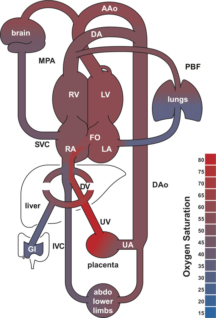
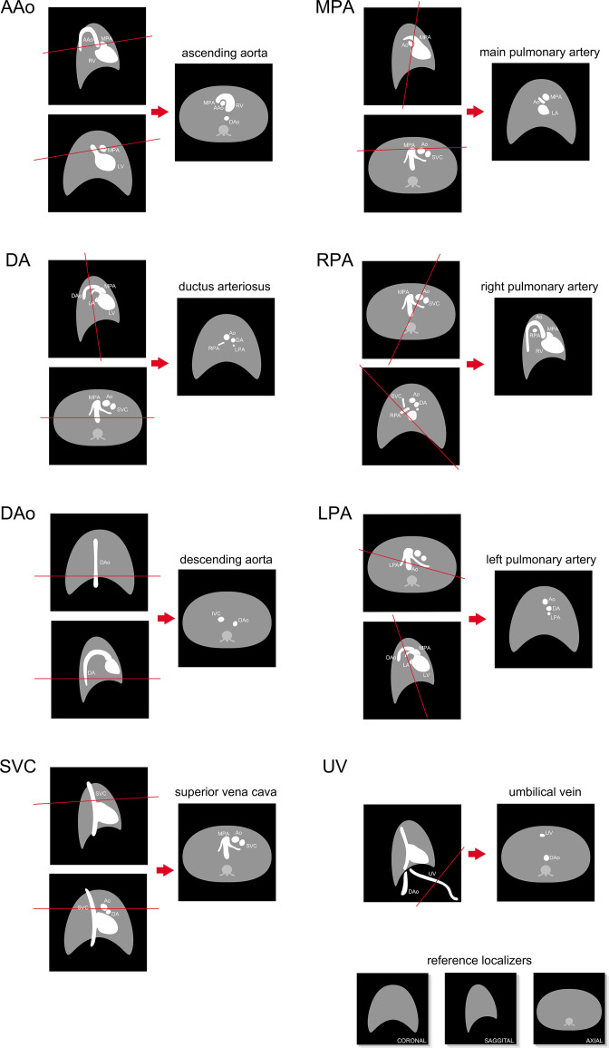
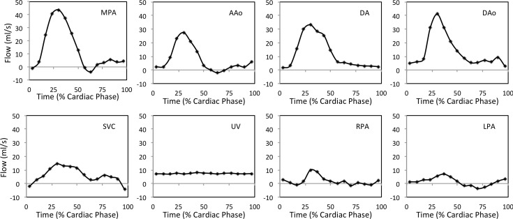
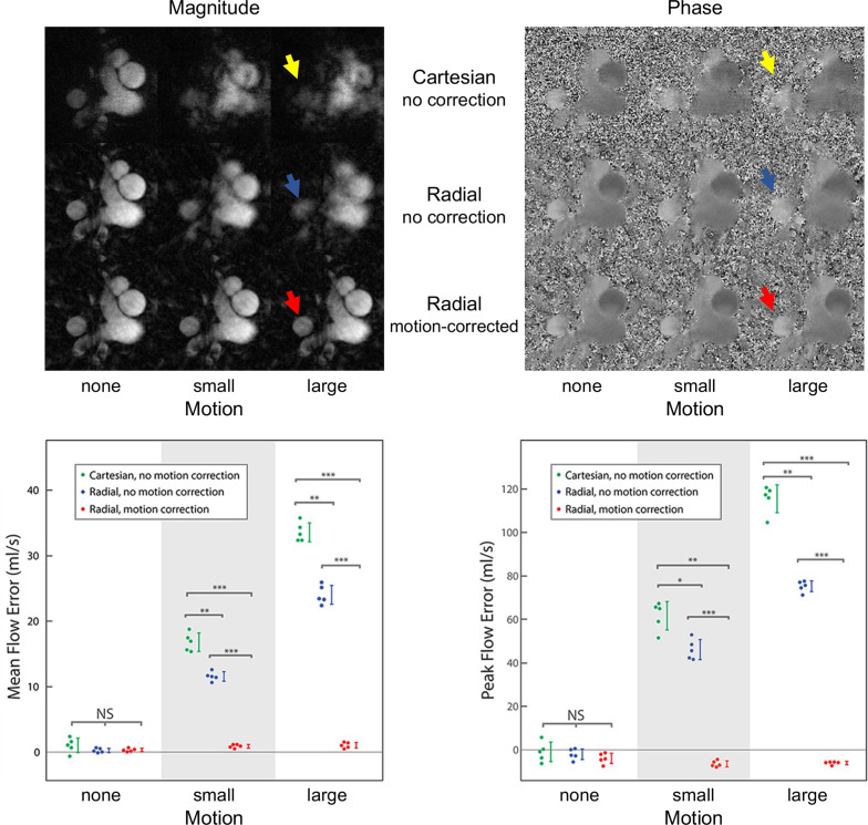
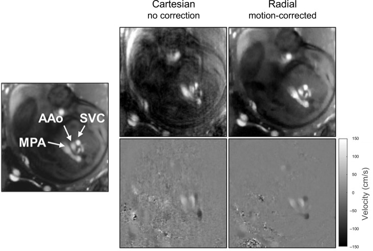
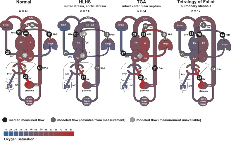
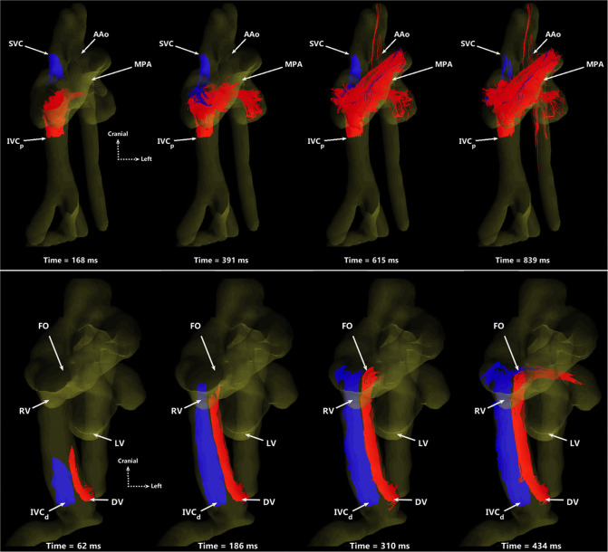
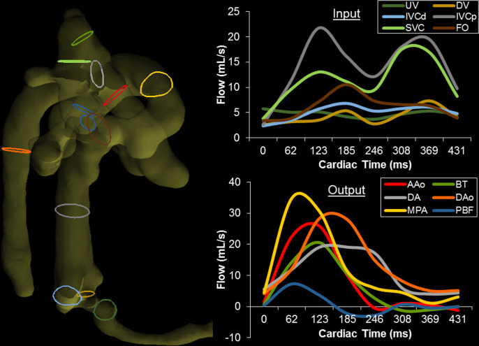
Fetal cardiac MRI is challenging due to fetal and maternal movements as well as the need for a reliable cardiac gating signal and high spatiotemporal resolution. Ongoing research and recent technical developments to address these challenges show the potential of MRI as an adjunct to ultrasound for the assessment of the fetal heart and great vessels. MRI measurements of blood flow have enabled the assessment of normal fetal circulation as well as conditions with disrupted circulations, such as congenital heart disease, along with associated organ underdevelopment and hemodynamic instability. This review provides details of the techniques used in fetal cardiovascular blood flow MRI, including single slice and volumetric imaging sequences, post-processing and analysis, along with a summary of applications in human studies and animal models.


Figures

Figure 1.
Figure 1.
Human fetal circulatory system diagram. The foramen ovale, ductus arteriosus and ductus venosus allow for mixing of the systemic and pulmonary circulations. These shunts are open during fetal development and close at birth. Shading indicates blood oxygen saturation measured in late gestational age fetuses using MR oximetry. AAo, ascending aorta; abdo, abdomen; DAo, decscending aorta; DA, ductus arteriosus, DV, ductus venosus; FO, foramen ovale; GI, gastrointestinal system; IVC, inferior vena cava; LA, left atrium; LV, left ventricle; MPA, main pulmonary artery; PBF, pulmonary blood flow; RA, right atrium; RV, right ventricle; UA, umbilical arteries; UV, umbilical vein. Adapted with permission from Ultrasound Obstet Gynecol 2021; doi: 10.1002/uog.23707.
Figure 2.
Figure 2.
Orientation of major fetal vessel cross-sectional slices in coronal, sagittal, and axial reference localizer images showing expected vessel arrangement in healthy human fetuses. AAo, ascending aorta; Ao, aorta; DAo, descending aorta; DA, ductus arteriosus; IVC, inferior vena cava; LPA, left pulmonary artery; LV, left ventricle; MPA, main pulmonary artery; RPA, right pulmonary artery; RV, right ventricle; SVC, superior vena cava; UV, umbilical vein. Figure and caption adapted with permission from MRI of Fetal and Maternal Diseases in Pregnancy. Springer, Cham. 2016. doi: 10.1007/978-3-319-21428-3_10.
Figure 3.
Figure 3.
Representative volume flow as a function of cardiac cycle in the major fetal vessels, measured by 2D PC MR in a healthy late-gestational age human fetus. AAo, ascending aorta; DAo, descending aorta; DA, ductus arteriosus; LPA, left pulmonary artery; MPA, main pulmonary artery; RPA, right pulmonary artery; SVC, superior vena cava; UV, umbilical vein. Reprinted under CC BY 2.0 from J Cardiovasc Magn Reson 2012;14(1):79. Caption adapted from original.
Figure 4.
Figure 4.
Impact of sampling and motion correction in simulated cine 2D phase contrast MR. (Top) Magnitude and velocity-sensitive phase contrast images of the upper great vessels based on adult reference data, simulated with Cartesian and golden angle radial k-space sampling under three motion states: none, small (±3 voxels displacement in each in-plane direction, approximately ±12% DAo diameter) and large (±5 voxels displacement,±20% DAo diameter). With large, uncorrected motion, identification of the DAo (arrows) boundary becomes more difficult but is ameliorated with motion correction. (Bottom) Mean and peak flow errors in the DAo. Vertical lines represent the standard deviation. Horizontal lines indicate significant differences. NS, not significant; *, p ≤ 0.05; **, p ≤ 10−4; ***, p ≤ 10−8; DAo, descending aorta. Figure and caption adapted with permission from J Magn Reson Imaging. 2021;53(2).
Figure 5.
Figure 5.
Cine phase contrast MR using Cartesian and motion-corrected golden angle radial sampling trajectories showing fetal three vessel view in systole. Mean and standard deviation of estimated in-plane displacement in the radial acquisition was 0.8 ± 0.5 mm with a maximum of 1.5 mm in images acquired with 1.3 mm resolution. Vessel labels are shown at left for reference. AAo, ascending aorta; MPA, main pulmonary artery; SVC, superior vena cava. Figure and caption adapted with permission from J Magn Reson Imaging. 2021;53(2).
Figure 6.
Figure 6.
Blood flow in the major vessels of normal controls and fetuses with cyanotic congenital heart disease. Scatter plots show (top) the distribution and group mean (bar) of flow normalized by fetal weight, and (bottom) group-wise difference plot with mean and 95% confidence interval for congenital heart disease subtypes compared to normal controls. Flows in congenital heart disease subtypes with confidence intervals that do not cross zero in the difference plot are significantly different from normal controls. Negative blood flows in the DA denote retrograde flow and left to right shunt in the FO. † Showing measured flows and, in cases with missing measurements, flows derived from other measurements. ‡ Showing derived flows exclusively. AAo, ascending aorta; CVO, combined ventricular output; DA, ductus arteriosus; DAo, descending aorta; EA, Ebstein’s anomaly; FO, foramen ovale; HLH, hypoplastic left heart; MPA, main pulmonary artery; PBF, pulmonary blood flow; SVC, superior vena cava; TA, tricuspid atresia; TGA, transposition of the great arteries; TOF, tetralogy of Fallot; UV, umbilical vein. Reprinted with permission from Ultrasound Obstet Gynecol 2021; doi: 10.1002/uog.23707. Caption adapted from original.
Figure 7.
Figure 7.
Distribution of blood flow in late gestation human fetuses with normal hearts and cyanotic congenital heart disease subtypes. Flows are shown as percentage of the combined ventricular output. Reported flows are based on median measured values subject to a constrained model ensuring continuity of flow volume throughout the fetal circulatory system. Negative values indicate retrograde flow or, in the case of the FO, left to right shunt. Shading indicates blood oxygen saturation measured by MR oximetry. AAo, ascending aorta; DA, ductus arteriosus; DAo, descending aorta; DV, ductus venosus; EA, Ebstein’s anomaly; HLHS, hypoplastic left heart syndrome; IVC, inferior vena cava; LA, left atrium; LV, left ventricle; MPA, main pulmonary artery; PBF, pulmonary blood flow; RA, right atrium; RV, right ventricle; SVC, superior vena cava; TGA, transposition of the great arteries; UV, umbilical vein; UA, umbilical arteries. Figure and caption adapted with permission from Ultrasound Obstet Gynecol 2021; doi: 10.1002/uog.23707.
Figure 8.
Figure 8.
Particle traces from 4D flow MR in fetal sheep. (Top) Oblique ventral view of SVC (blue) and IVCp (red) return to the right side-of the heart, shown at four time points over two cardiac cycles. The MPA receives blood from both vessels, serving as a conduit to the lungs and lower body. The AAo only receives IVCp blood via right-to-left atrial shunting. (Bottom) Ventral view showing preferential delivery of DV blood to the left side of the heart. DV (red) and IVCd (blue) particles are shown at four time points over one cardiac cycle. The two streams remain well-separated, with blood from the DV primarily entering the left heart through the FO while IVCd remains in the right heart. SVC, superior vena cava; IVCp, proximal inferior vena cava; AAo, ascending aorta; MPA, main pulmonary artery; DV, ductus venosus; IVCd, distal inferior vena cava; FO, foramen ovale; RV, right ventricle; LV, left ventricle. Figure and caption adapted from J Cardiovasc Magn Reson. 2019; 21(1) under CC BY 4.0.
Figure 9.
Figure 9.
Right lateral view of the segmented fetal sheep vasculature for quantitation of blood flow volumes. Color-coded contours indicate locations of corresponding flow measurements at right. (Right) Blood flow vs cardiac time at all measurement locations, separated as (top) input to and (bottom) output from the heart. Cardiac inputs from the DV, IVCd, IVCp, and SVC display typical biphasic nature with greater pulsatility proximal to the heart, whereas UV flow appears nearly constant. Note UV flow was measured downstream from the junction of the typical two UVs found in fetal sheep. Outputs from the MPA, DA, AAo, BT, and DAo display large systolic peaks and low diastolic flows. BT and PBF display retrograde diastolic flow, likely indicative of runoff to the placenta through the aortic isthmus and DA. UV, umbilical vein; DV, ductus venosus; IVCd, distal inferior vena cava; IVCp, proximal inferior vena cava; SVC, superior vena cava; FO, foramen ovale; AAo, ascending aorta; BT, brachiocephalic trunk; DA, ductus arteriosus; DAo, descending aorta; MPA, main pulmonary artery; PBF, combined pulmonary blood flow. Reprinted from J Cardiovasc Magn Reson. 2019; 21(1) under CC BY 4.0. Caption adapted from original.

References

    1. Yagel S, Silverman NH, Gembruch U, eds. Circulation in the normal fetus and cardiovascular adaptations to birth. Fetal Cardiology 2019; 119–37. Available from: 10.1201/9780429461118-16 - DOI
    1. Rudolph A. Fetal Cardiology. 3rd ed. CRC Press; 2019., pp.708–22. Available from: 10.1201/9780429461118-61 - DOI
    1. Peyvandi S, Donofrio MT. Circulatory changes and cerebral blood flow and oxygenation during transition in newborns with congenital heart disease. Semin Pediatr Neurol 2018; 28: 38–47. doi: 10.1016/j.spen.2018.05.005 - DOI - PubMed
    1. Donofrio MT, Moon-Grady AJ, Hornberger LK, Copel JA, Sklansky MS, Abuhamad A, et al. . Diagnosis and treatment of fetal cardiac disease: A scientific statement from the american heart association. Circulation 2014; 129: 2183–2242. doi: 10.1161/01.cir.0000437597.44550.5d - DOI - PubMed
    1. Gill RW. Measurement of blood flow by ultrasound: accuracy and sources of error. Ultrasound Med Biol 1985; 11: 625–41. doi: 10.1016/0301-5629(85)90035-3 - DOI - PubMed