. 2022 Jun 10;7(72):eabl9330.
doi: 10.1126/sciimmunol.abl9330. Epub 2022 Jun 10.

ATR-mediated CD47 and PD-L1 up-regulation restricts radiotherapy-induced immune priming and abscopal responses in colorectal cancer

Affiliations

ATR-mediated CD47 and PD-L1 up-regulation restricts radiotherapy-induced immune priming and abscopal responses in colorectal cancer

Rodney Cheng-En Hsieh et al. Sci Immunol. .

Abstract

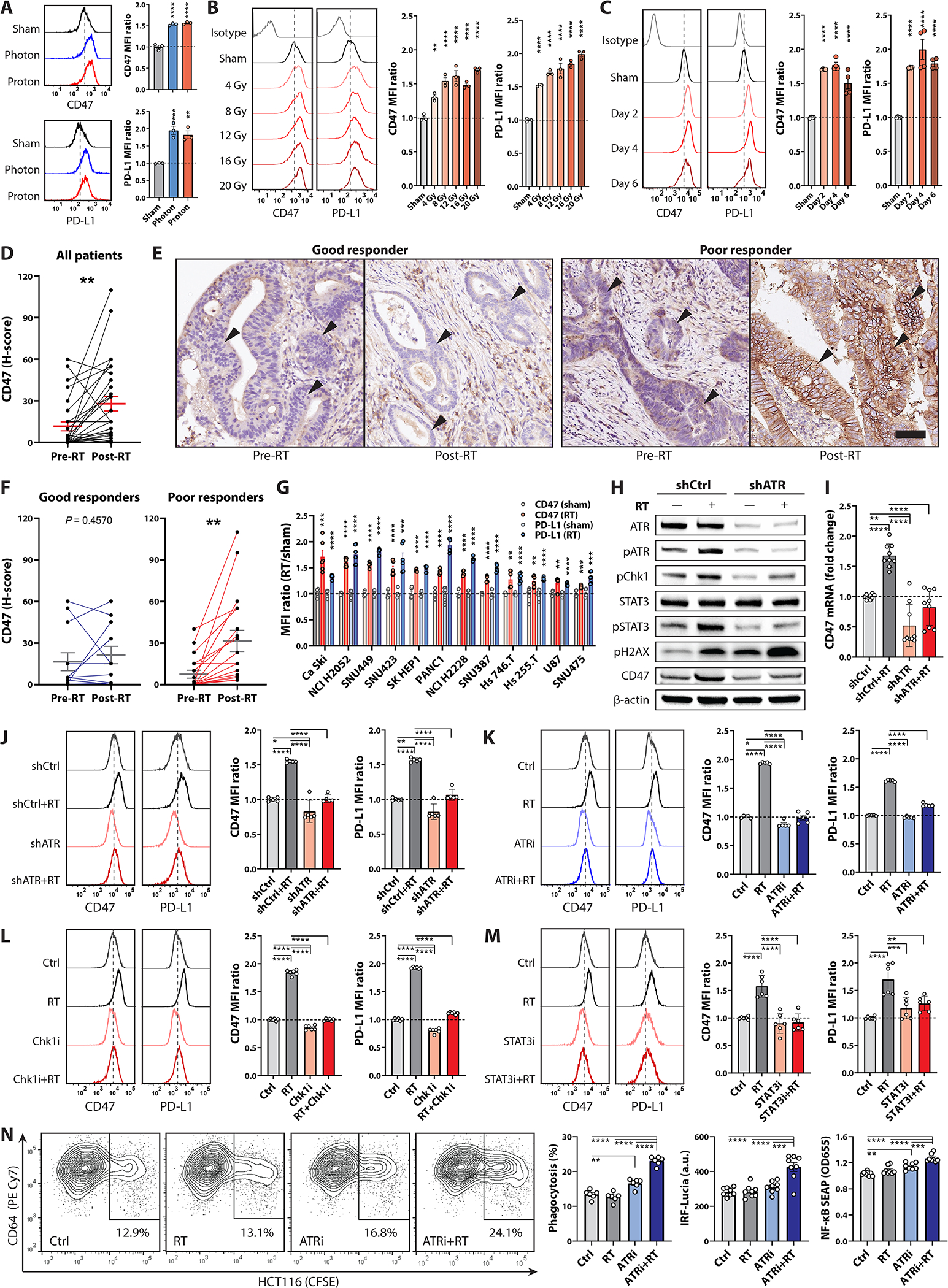
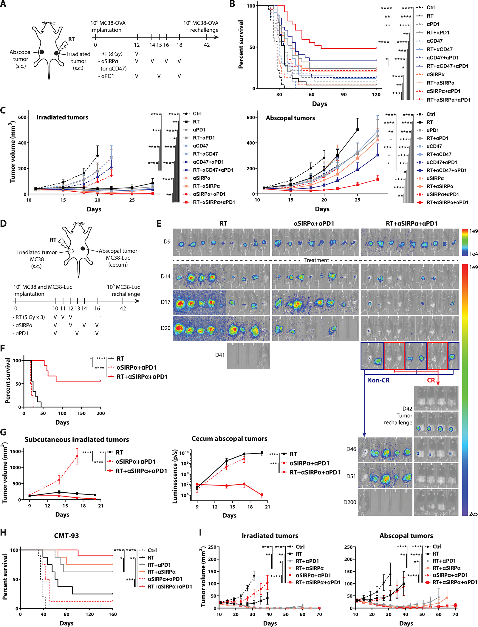
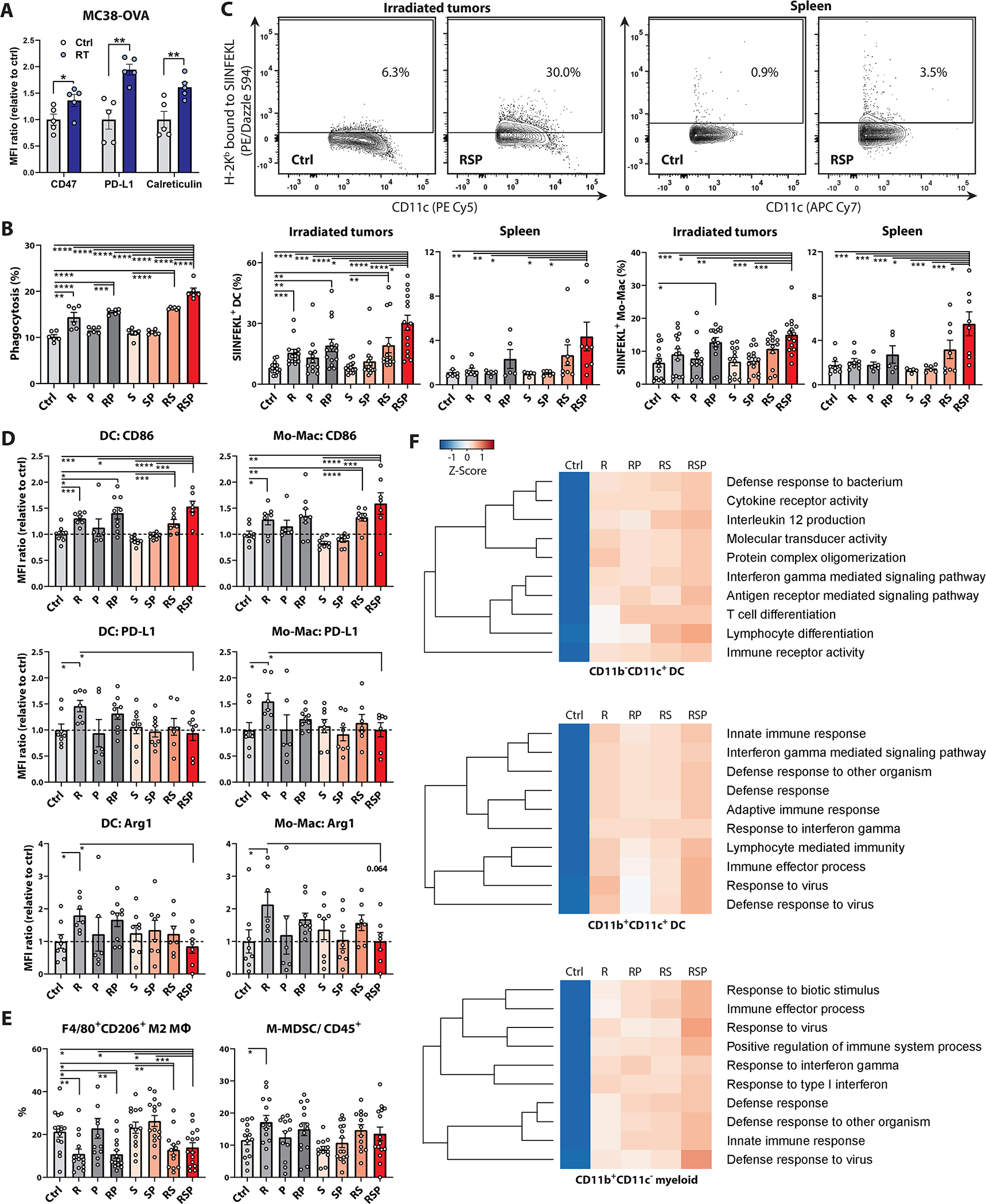
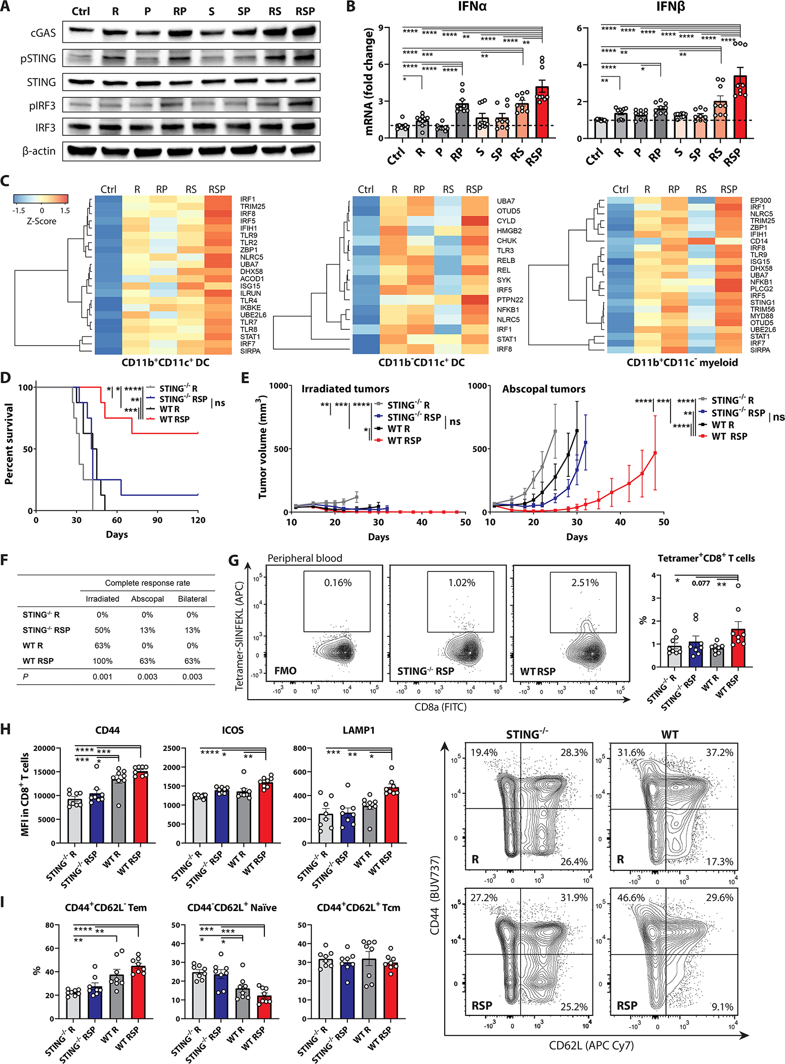
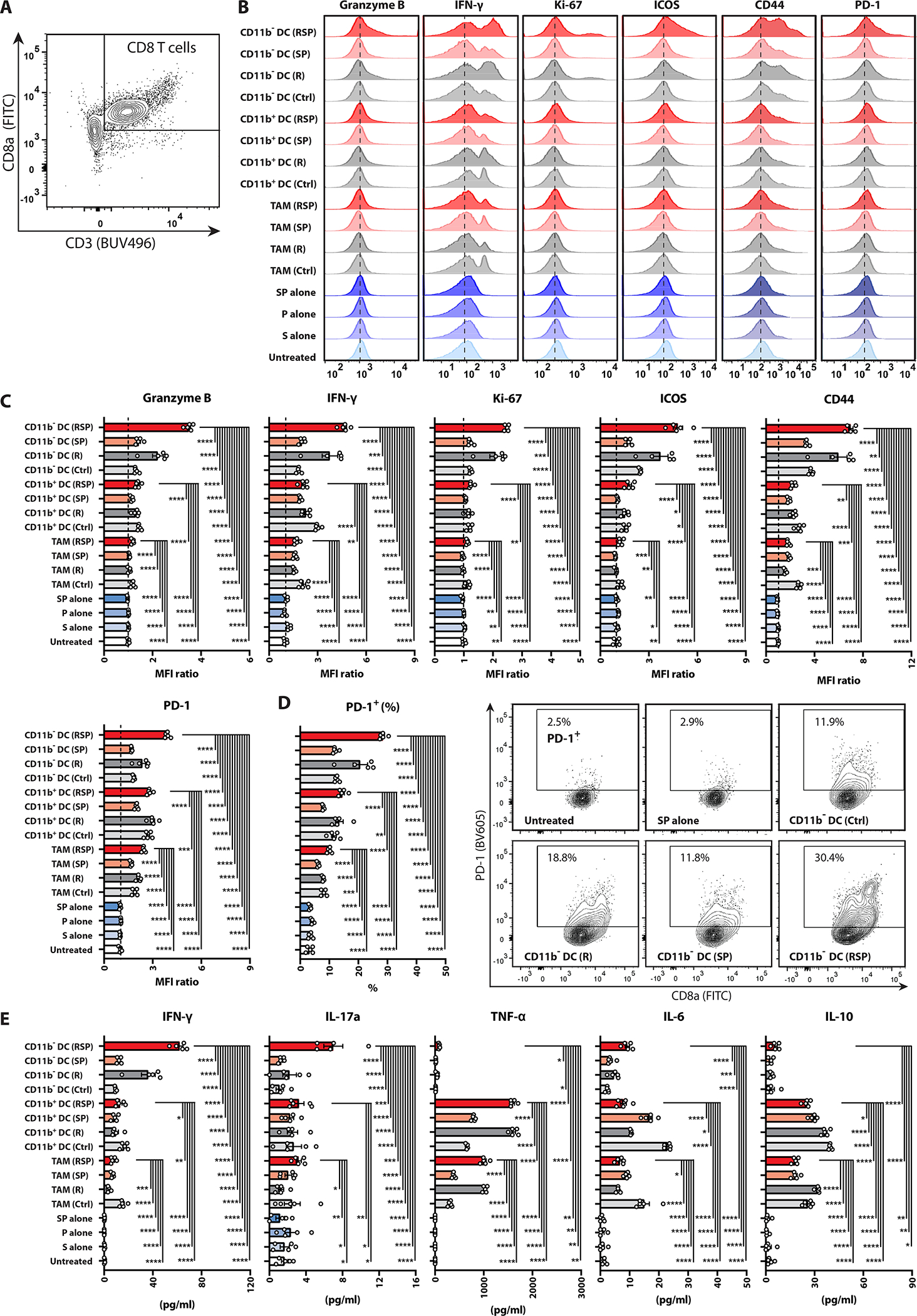
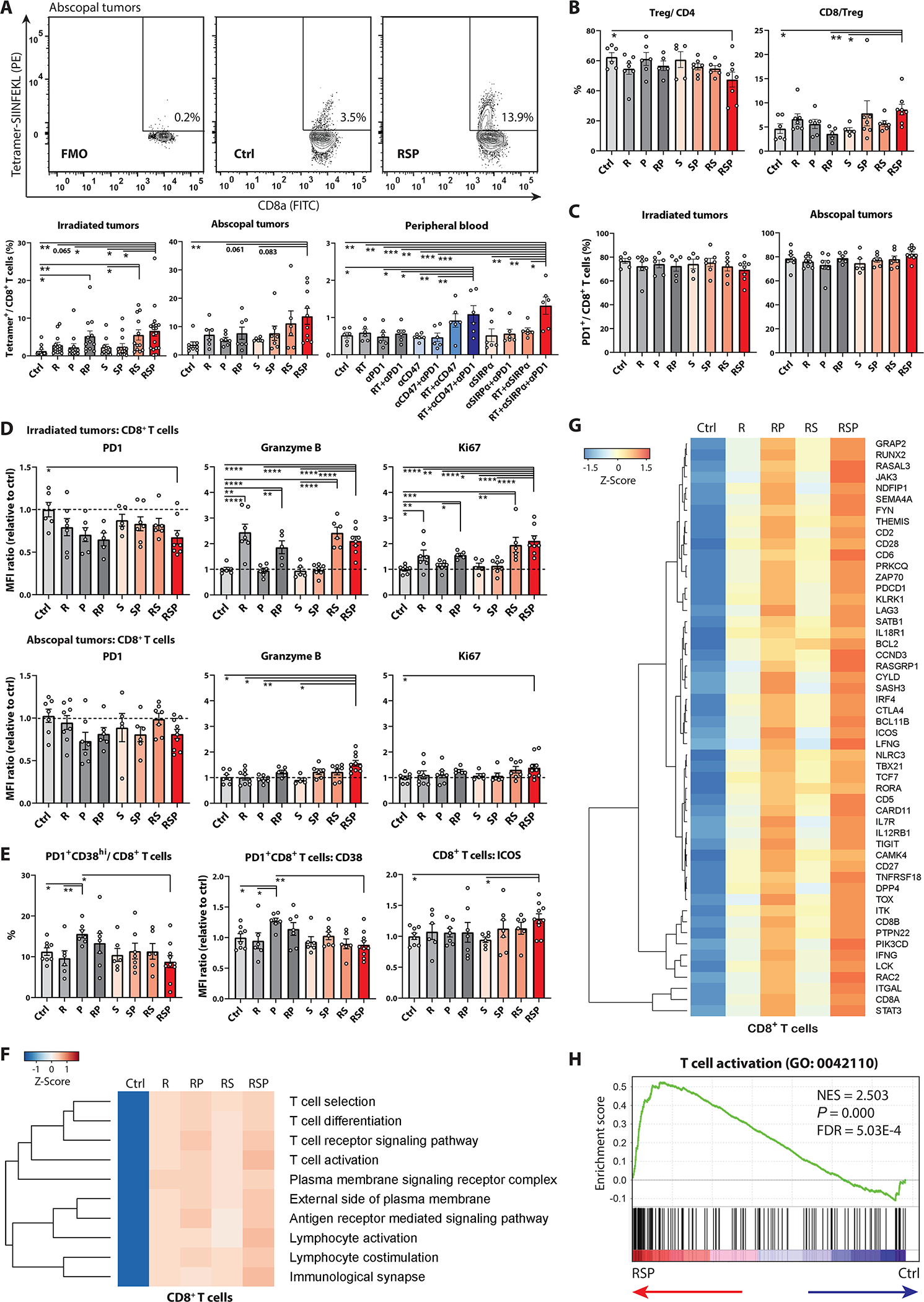
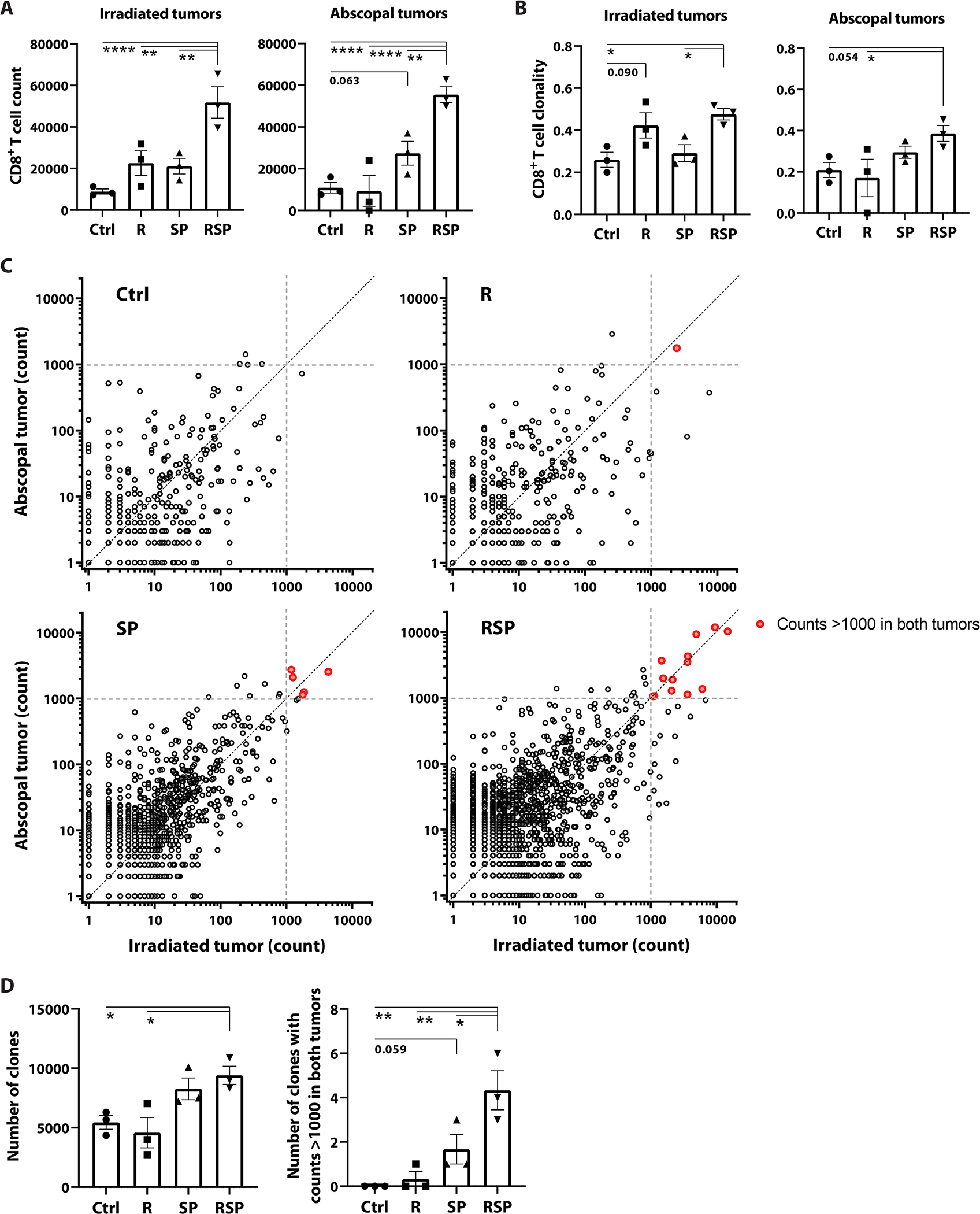
Radiotherapy (RT) of colorectal cancer (CRC) can prime adaptive immunity against tumor-associated antigen (TAA)-expressing CRC cells systemically. However, abscopal tumor remissions are extremely rare, and the postirradiation immune escape mechanisms in CRC remain elusive. Here, we found that irradiated CRC cells used ATR-mediated DNA repair signaling pathway to up-regulate both CD47 and PD-L1, which through engagement of SIRPα and PD-1, respectively, prevented phagocytosis by antigen-presenting cells and thereby limited TAA cross-presentation and innate immune activation. This postirradiation CD47 and PD-L1 up-regulation was observed across various human solid tumor cells. Concordantly, rectal cancer patients with poor responses to neoadjuvant RT exhibited significantly elevated postirradiation CD47 levels. The combination of RT, anti-SIRPα, and anti-PD-1 reversed adaptive immune resistance and drove efficient TAA cross-presentation, resulting in robust TAA-specific CD8 T cell priming, functional activation of T effectors, and increased T cell clonality and clonal diversity. We observed significantly higher complete response rates to RT/anti-SIRPα/anti-PD-1 in both irradiated and abscopal tumors and prolonged survival in three distinct murine CRC models, including a cecal orthotopic model. The efficacy of triple combination therapy was STING dependent as knockout animals lost most benefit of adding anti-SIRPα and anti-PD-1 to RT. Despite activation across the myeloid stroma, the enhanced dendritic cell function accounts for most improvements in CD8 T cell priming. These data suggest ATR-mediated CD47 and PD-L1 up-regulation as a key mechanism restraining radiation-induced immune priming. RT combined with SIRPα and PD-1 blockade promotes robust antitumor immune priming, leading to systemic tumor regressions.


Conflict of interest statement

Conflict of interest:

M.A.C. reports grants and personal fees from ImmunoGenesis, Inc., personal fees from Alligator Bioscience, Inc., personal fees from ImmunOS, Inc., grants and personal fees from ImmunoMet, Inc., personal fees from Oncoresponse, Inc., personal fees from Pieris, Inc., personal fees from Nurix, Inc., personal fees from Aptevo, Inc., personal fees from Servier, Inc., personal fees from Kineta, Inc., personal fees from Salarius, Inc., personal fees from Xencor, Inc., personal fees from Agenus, Inc., personal fees from Mereo, Inc., personal fees from Amunix, Inc., personal fees from Adagene, Inc., outside the submitted work; in addition, M.A.C. has a patent (US 9,868,961 B2) Methods and Composition for Localized Secretion of Anti-CTLA-4 Antibodies with royalties paid to multiple licensees, a patent (PCT/US2019/022295) Dual specificity antibodies which bind both PD-L1 and PD-L2 and prevent their binding to PD-1 with royalties paid to ImmunoGenesis, Inc., and a patent (#17/065,304) Cyclic Dinucleotides as Agonists of Stimulator of Interferon Gene Dependent Signaling licensed to ImmunoGenesis, Inc.. C.M.T. is an inventor on the patent (#16/766,025) held by MD Anderson Cancer Center that covers radioprotection of the gastrointestinal tract with oral amifostine. C.M.T. is on the medical advisory board of Accuray and is a paid consultant for Xerient Pharma and Phebra Pty, Ltd. The remaining authors declare that they have no competing interests.

Figures

Figure 1 |
Figure 1 |. Irradiated CRC cells upregulate CD47 and PD-L1 to evade phagocytosis through ATR-Chk1-STAT3 signaling.
Mean fluorescent intensity (MFI) of CD47 and PD-L1 on HCT116 cells (A) 48 hours after photon (8 Gy) or proton (8 CGE) irradiation, (B) 48 hours after 4–20 Gy or sham irradiation and (C) 2–6 days after 8-Gy or sham irradiation determined via flow cytometry (n = 3–4). D-F, Histoscores (H-scores; D and F) and (E) representative images of CD47 immunohistochemistry surface staining on CRC cells (arrowheads) in pretreatment biopsies versus post-irradiation surgical specimens in 30 rectal cancer patients who underwent short-course neoadjuvant radiotherapy (RT, 5 Gy × 5 Fr) followed by en-bloc resection. Bars on H-scores, mean ± standard errors (paired t-test); good responders, Mandard tumor regression grade (TRG) 2–3; poor responders, TRG 4–5; scale bar, 100 μm. G, CD47 and PD-L1 MFI in various human solid cancer cells after 8-Gy irradiation (n = 6). H-J, Expression of CD47 and PD-L1 on scrambled shRNA control (shCtrl) versus ATR shRNA-knockdown (shATR) HCT116 cells treated +/− 8 Gy radiotherapy. ATR knockdown was assessed by (H) immunoblotting 24 hours after 8-Gy or sham irradiation (n = 3 biological replicates). I, Transcriptional levels of CD47 in shCtrl or shATR clones 24 hours after 8-Gy or sham irradiation (n = 9). J, Histograms and MFI of CD47 and PD-L1 on shCtrl- or shATR-HCT116 cells 48 hours after 8-Gy or sham irradiation (n = 5). K-M, Histograms and MFI of CD47 and PD-L1 on HCT116 cells 48 hours after 8-Gy or sham irradiation +/− (K) ATRi (300 nM VE822), (L) Chk1i (300 nM UCN-01), or (M) STAT3i (100 μM NSC74859; n = 5–6). N, Phagocytosis assays using differentiated THP1-Dual cells, which expresses IRF and NF-κB reporters, co-cultured with CFSE-labeled HCT116 cells +/− RT and ATR inhibition (300 nM VE822, n = 6–9). Phagocytosis is expressed as the percentage of CFSE+ cells in CD64+ THP1-Dual cells. *P <0.05, **P <0.01, ***P <0.001, ****P <0.0001.
Figure 2 |
Figure 2 |. RT coupled with phagocytosis checkpoint blockade clears both irradiated and abscopal tumors, inducing antitumor immune memory and prolonged survival.
Phagocytosis checkpoint blocking antibodies –anti-CD47 (αCD47; 400 μg/dose/mouse, 4 doses), anti-SIRPα (αSIRPα; 400 μg/dose/mouse, 4 doses), and anti-PD-1 (αPD1; 250 μg/dose/mouse, 3 doses) were administered intraperitoneally in C57BL/6J mice. Focal tumor irradiation was performed using cone-beam computed tomography-guided radiotherapy with 1.5 cm beamwidth. A, Schematic diagram in mice implanted with bilateral MC38-ovalbumin (OVA) tumors on flank subcutaneous regions and treated +/− (1) 8-Gy irradiation to unilateral tumors, (2) anti-CD47 or anti-SIRPα antibody, and/or (3) anti-PD-1 antibody (n = 23–27). MC38-OVA tumor rechallenge was performed on day 42 in mice with bilateral complete responses (CR). B, Survival and (C) tumor growth curves for panel A. D, Schematic diagram in mice bearing right flank subcutaneous wild-type MC38 and cecal orthotopic MC38-luciferase (MC38-Luc) tumors treated with either focal RT (to right flank tumors) with 5 Gy for 3 fractions, anti-SIRPα and anti-PD-1 antibodies, or both (n = 8–9). Rechallenge of MC38-Luc cells on the left flank was performed on day 42 in mice with CR of both tumors. E, Bioluminescent images, (F) survival, and (G) tumor growth kinetics for panel D. H, Survival and (I) tumor growth curves in mice implanted with bilateral CMT-93 tumors treated +/− focal RT (day 12 and 13) with 5 Gy for 2 fractions to unilateral tumors, anti-SIRPα (day 12, 14, 16, and 18), and/or anti-PD-1 (day 12, 15, and 18) (n = 8–10). Cured mice were rechallenged with CMT-93 tumor cells on day 45. *P <0.05, **P <0.01, ***P <0.001, ****P <0.0001.
Figure 3 |
Figure 3 |. RT/anti-SIRPα/anti-PD-1 (RSP) promotes tumor antigen cross-priming, APC mobilization, and myeloid compartment activation.
A-E, MFI and percentages were measured using flow cytometry. A, MFI of CD47, PD-L1, and calreticulin on MC38-OVA cells extracted from tumor-bearing mice 48 hours after 8-Gy or sham irradiation (n = 5). B, Phagocytosis assay of MC38-OVA cells using bone marrow-derived dendritic cells (BM-DCs). MC38-OVA cells were labeled with CFSE 48 hours after 8-Gy or sham radiotherapy (R). BM-DCs were pretreated +/− anti-SIRPα (20 μg/ml) and anti-PD-1 (12.5 μg/ml) for 30 minutes and co-cultured with CFSE-labeled MC38-OVA cells under the same antibody condition for 4 hours at 37 °C. Phagocytosis is expressed as the percentage of CFSE+ cells in CD11c+ BM-DCs (n = 6). C-F, Leukocytes in irradiated tumors and spleens were harvested and dispersed into single-cell suspensions on day 17 from MC38-OVA tumor-bearing mice treated +/− 8-Gy radiotherapy (R; day 12), anti-SIRPα (S; day 12, 14, and 16), and/or anti-PD-1 (P; day 12 and 15). C, Percentages of H-2Kb-SIINFEKL+ cross-presenting DC, monocytes, and macrophages (Mo-Macs) in irradiated tumors and spleen. D, MFI of CD86, PD-L1, and Arginase1 (Arg1) in DCs and Mo-Macs. E, Percentages of CD206+ M2-macrophages in CD11b+F4/80+ cells and the percentages of CD11b+Ly6GLy6Chi monocytic MDSCs (M-MDSCs) in CD45+ cells. (C-E, n = 5 to 15). F, Hierarchical clusters of top 10 enriched Gene Ontology pathways in the transcriptomic analyses of CD11bCD11c+, CD11b+CD11c+, and CD11b+CD11c APCs which were isolated from irradiated tumors using cell sorters. The tumor-infiltrating leukocytes were pooled from 10 tumor-bearing mice for each group to increase RNA yield and sample representativeness. *P <0.05, **P <0.01, ***P <0.001, ****P <0.0001.
Figure 4 |
Figure 4 |. RT/anti-SIRPα/anti-PD-1 (RSP)-induced immune priming is mediated by host cGAS-STING.
A-B, BM-DCs were pretreated +/− anti-SIRPα (S; 20 μg/ml) and anti-PD-1 (P; 12.5 μg/ml) for 30 minutes and co-cultured with 8-Gy (R) or sham irradiated MC38-OVA cells (tumor: BM-DC = 1:3) under the same antibody condition for 1 hours at 37 °C. BM-DCs were isolated using CD11c MicroBeads for (A) immunoblotting of cGAS, pSTING, STING, pIRF3, IRF3, and β-actin (n = 3 biological replicates) and (B) RT-PCR of interferon (IFN)-α and -β (n = 9). C, Differentially expressed gene analyses in type I IFN production (GO: 0032606)-related genes in different treatment groups across CD11b+CD11c+, CD11bCD11c+, and CD11b+CD11c APCs which were isolated from irradiated tumors using cell sorters. Each RNA sample was extracted from the pooled tumor-infiltrating APCs from 10 MC38-OVA tumor-bearing mice.D, Survival, (E) tumor control curves, and (F) complete response rates in wild-type (WT) versus STING-deficient (STING−/−) mice bearing bilateral MC38-OVA tumors treated with 8-Gy radiotherapy (R) to unilateral tumors +/− anti-SIRPα (S; day 12, 14, 16, and 18) and anti-PD-1 (P; day 12, 15, and 18. G-I, The CD8 T cells in peripheral blood were harvested on day 19 from STING−/− versus WT mice treated with RT alone or RT/anti-SIRPα/anti-PD-1 for flow cytometry analyses. G, Frequencies of H2-Kb-SIINFEKL-tetramer+ CD8 T cells. H, MFI of CD44, ICOS, and surface LAMP1 on circulating CD8 T cells. I, Frequencies of naïve (CD44CD62L+), effector memory (CD44+CD62L, Tem), and central memory (CD44+CD62L+, Tcm) CD8 T cells in the periphery. (D-I, n = 8) *P <0.05, **P <0.01, ***P <0.001, ****P <0.0001.
Figure 5 |
Figure 5 |. RT/anti-SIRPα/anti-PD-1 (RSP)-induced TAA-specific CD8 T cell cross-priming is predominantly driven by CD11bCD11c+ DCs.
The Ex vivo TAA-specific CD8 T cell cross-priming assay was performed by co-culturing unstimulated splenic OT-1 cells with tumor-infiltrating CD11bCD11c+ DCs (mostly cDC1), CD11b+CD11c+ DCs, or CD11b+CD11c F4/80+ TAMs (OT-1: APCs = 2:1), which were harvested from MC38-OVA tumor-bearing mice treated +/− ipsilateral 8 Gy radiotherapy (R; day 12), anti-SIRPα (S; day 12, 14, 16), and/or anti-PD-1 (P; day 12, 15) on day 17, or with anti-SIRPα (20 μg/ml) and/or anti-PD-1 (12.5 μg/mL) antibodies alone (blue bars) for 48 hours. A, Gating strategy for CD8+ OT-1 T cells. B, Histograms and (C) MFI of granzyme B, IFN-γ, Ki-67, ICOS, CD44, and PD-1 in OT-1 cells (relative to untreated controls). D, Percentages and contour plots of PD-1+ OT-1 cells E, Concentrations of secreted IFN-γ, IL-17a, TNF-α, IL-6, and IL-10 in the supernatant of OT-1 cells co-cultured with different APCs or antibody conditions measured by cytometric bead array. *P <0.05, **P <0.01, ***P <0.001, ****P <0.0001.
Figure 6 |
Figure 6 |. RT/anti-SIRPα/anti-PD-1 (RSP) promotes anti-tumor T cell activation.
A, T cells in irradiated tumors, abscopal tumors, and peripheral blood were harvested on day 17, 19, and 19, respectively, from mice bearing bilateral MC38-OVA tumors treated +/− ipsilateral 8-Gy radiotherapy (R; day 12), anti-SIRPα (S; day 12, 14, 16, 18), and/or anti-PD-1 (P; day 12, 15, 18) for flow cytometry analyses. A, Frequencies of H-2Kb-SIINFEKL-tetramer+ CD8 T cells in irradiated and abscopal tumors as well as the peripheral blood. Contour plots depict the percentages of H-2Kb-SIINFEKL-tetramer+ cells in CD8 T lymphocytes in abscopal tumors. B-D, T cells in irradiated and abscopal tumors were harvested on day 17 from mice bearing bilateral MC38-OVA tumors treated +/− ipsilateral 8-Gy radiotherapy (R; day 12), anti-SIRPα (S; day 12, 14, 16), and/or anti-PD-1 (P; day 12, 15). B, Frequencies of FoxP3+ Tregs and CD8/Treg ratios in irradiated tumors. C, Percentages of PD1+ CD8 T cells in irradiated and abscopal tumors. D, MFI of PD-1, Granzyme B, and Ki-67 in CD8 T cells in irradiated (upper panels) and abscopal (lower panels) tumors. E, Percentages of PD1+CD38hi subprimed CD8 T cells and MFI of CD38 in PD1+ CD8 T cells and ICOS in CD8 T cells in abscopal tumors. The treatment condition is the same as A. (A-E, n = 5 to 18) F-H, CD8 T cells were isolated from irradiated tumors on day 17 from mice bearing bilateral MC38-OVA tumors treated +/− ipsilateral 8-Gy radiotherapy (R; day 12), anti-SIRPα (S; day 12, 14, and 16) and/or anti-PD-1 (P; day 12 and 15) using cell sorters for RNA-seq analyses. F, Hierarchical clusters of top 10 enriched Gene Ontology pathways in tumor-infiltrating CD8 T cells. G, Differentially expressed gene analyses in T cell activation-associated genes (GO: 0042110) in CD8 T cells across different treatment groups. H, Leading edge plot in the T cell activation geneset in the RT/anti-SIRPα/anti-PD-1 group. (F-H, Each RNA sample was extracted from the pooled CD8 T cells from 10 MC38-OVA tumor-bearing mice.) *P <0.05, **P <0.01, ***P <0.001, ****P <0.0001.
Figure 7 |
Figure 7 |. RT/anti-SIRPα/anti-PD-1 (RSP) expands T cell clonality and clonal diversity.
CD8 T cells in irradiated and abscopal tumors were isolated on day 19 from mice bearing bilateral MC38-OVA tumors treated +/− ipsilateral 8-Gy radiotherapy (R; day 12), anti-SIRPα (S; day 12, 14, 16, 18), and/or anti-PD-1 (P; day 12, 15, 18) using cell sorters. Genomic DNA was extracted for high-throughput sequencing of TCRβ CDR3 to characterize the landscape of tumor-infiltrating CD8 T cell repertoire. (n = 3). A, Tumor-infiltrating CD8 T cell counts (determined by the sum of template counts for all productive rearrangements in each sample) in irradiated and abscopal tumors. B, CD8 T cell clonality indices in irradiated and abscopal tumors. C, Scatterplots of clonal abundance in irradiated versus abscopal tumors. Each dot represents one unique TCR clone; red dots indicate the expanded clones with cell counts >1000 in both tumors. Each panel comprises three independent samples. D, The total number of unique TCR clones (left) and the number of expanded clones with counts >1000 in both tumors (right). *P <0.05, **P <0.01, ***P <0.001, ****P <0.0001.

References

    1. Sung H, Ferlay J, Siegel RL, Laversanne M, Soerjomataram I, Jemal A, Bray F, Global Cancer Statistics 2020: GLOBOCAN Estimates of Incidence and Mortality Worldwide for 36 Cancers in 185 Countries. CA: A Cancer Journal for Clinicians 71, 209–249 (2021). - PubMed
    1. Tam SY, Wu VWC, A Review on the Special Radiotherapy Techniques of Colorectal Cancer. Front Oncol 9, 208–208 (2019). - PMC - PubMed
    1. Bach SP, Gilbert A, Brock K, Korsgen S, Geh I, Hill J, Gill T, Hainsworth P, Tutton MG, Khan J, Robinson J, Steward M, Cunningham C, Levy B, Beveridge A, Handley K, Kaur M, Marchevsky N, Magill L, Russell A, Quirke P, West NP, Sebag-Montefiore D, Brown G, Antonio P, Vince A, Hilken N, Sidile C, Wilcockson A, Peto R, Crosby T, Moran B, Olliff J, Ashok K, Slawik S, Smethurst A, Sripadam R, Tagore V, Terlizzo M, Philip B, Davies R, Dodd S, Essapen S, Nisar P, Stewart A, Trickett J, Ashish B, Billings P, Chandran P, Corr C, Favill E, Gollins S, Marsh P, Maw A, Neupane R, Rajagopal R, Cooper R, Griffith J, Hatfield P, Lowe A, Ostrowski J, Robinson J, Simpson R, Adams R, Bleehen R, Davies M, Morgan M, Boone D, Lacey N, Seddon I, Sizer B, Stunell H, Wu S, Hadaki M, Blunt D, Cleator S, Darzi A, Goldin R, Ziprin P, Dobson M, Pitt M, Susnerwala S, Williamson D, Howarth G, Lee S, Wright P, Hoare T, Horgan A, McDonald F, Needham S, Scott J, Simmons T, Biswas D, Hernon J, Kapur G, Kapur S, Sington J, Speakman C, Stebbings W, Williams S, Adusumalli M, Agarwal A, Borowski D, Garg D, Gill T, Hegab M, Hobday C, Rao V, Shrimankar J, Tabaqchali M, Wilson D, Jones O, Mortensen N, Slater A, Szuts A, Wang L, Warren B, Weaver A, Ahmad M, Alexander J, Flubacher M, Tarver D, Baluch S, Beable R, Cowlishaw D, Higginson A, Vogiatzis P, Cruickshank N, Joy H, Peake D, Zanetto U, Saunders M, Sun-Myint A, Sripadam R, Cooper R, Hatfield P, Teo M, Allan A, Geh I, Glaholm J, Goldstein M, Hejmadi R, Langman G, Morton D, Nelson C, Tattersall D, Falk S, Longman R, Roach H, Shabbir J, Shelley-Fraser G, Thomas M, Cripps N, Haba Y, Harris G, Hookway M, Simson J, Skull A, Umar T, Radical surgery versus organ preservation via short-course radiotherapy followed by transanal endoscopic microsurgery for early-stage rectal cancer (TREC): a randomised, open-label feasibility study. The Lancet Gastroenterology & Hepatology 6, 92–105 (2021). - PMC - PubMed
    1. van der Valk MJM, Marijnen CAM, van Etten B, Dijkstra EA, Hilling DE, Kranenbarg EM-K, Putter H, Roodvoets AGH, Bahadoer RR, Fokstuen T, ten Tije AJ, Capdevila J, Hendriks MP, Edhemovic I, Cervantes AMR, de Groot DJA, Nilsson PJ, Glimelius B, van de Velde CJH, Hospers GAP, Compliance and tolerability of short-course radiotherapy followed by preoperative chemotherapy and surgery for high-risk rectal cancer – Results of the international randomized RAPIDO-trial. Radiotherapy and Oncology 147, 75–83 (2020). - PubMed
    1. Wennerberg E, Lhuillier C, Vanpouille-Box C, Pilones KA, García-Martínez E, Rudqvist N-P, Formenti SC, Demaria S, Barriers to Radiation-Induced In Situ Tumor Vaccination. Frontiers in Immunology 8, (2017). - PMC - PubMed

Publication types