. 2023 Jan 17;62(2):419-428.
doi: 10.1021/acs.biochem.2c00168. Epub 2022 Jun 10.

Rationally Guided Improvement of NOV1 Dioxygenase for the Conversion of Lignin-Derived Isoeugenol to Vanillin

Affiliations

Rationally Guided Improvement of NOV1 Dioxygenase for the Conversion of Lignin-Derived Isoeugenol to Vanillin

Mario De Simone et al. Biochemistry. .

Abstract

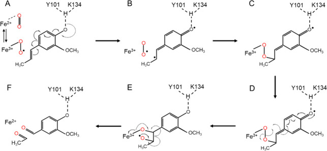
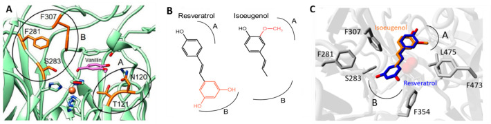
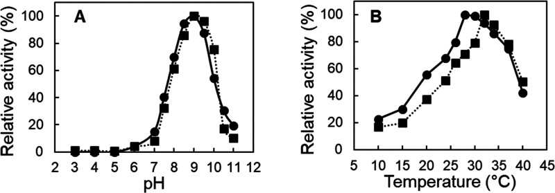
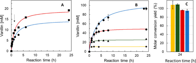
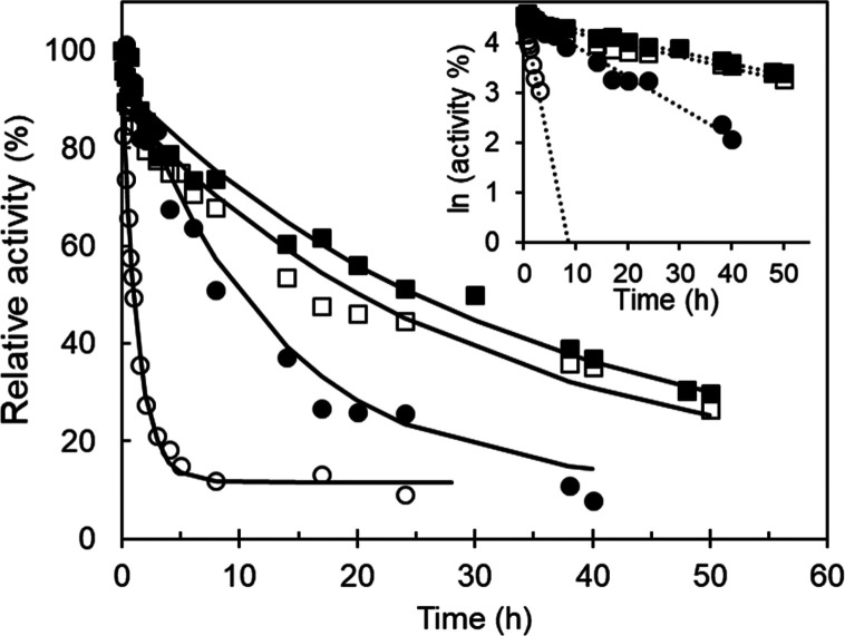
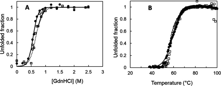
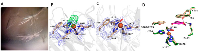
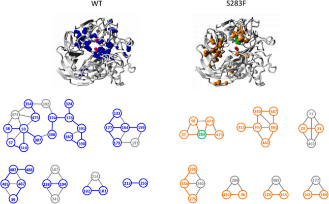
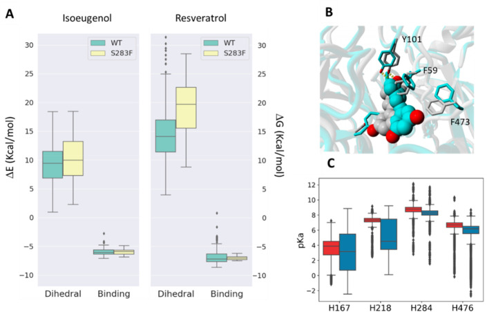
Biocatalysis is a key tool in both green chemistry and biorefinery fields. NOV1 is a dioxygenase that catalyzes the one-step, coenzyme-free oxidation of isoeugenol into vanillin and holds enormous biotechnological potential for the complete valorization of lignin as a sustainable starting material for biobased chemicals, polymers, and materials. This study integrates computational, kinetic, structural, and biophysical approaches to characterize a new NOV1 variant featuring improved activity and stability compared to those of the wild type. The S283F replacement results in a 2-fold increased turnover rate (kcat) for isoeugenol and a 4-fold higher catalytic efficiency (kcat/Km) for molecular oxygen compared to those of the wild type. Furthermore, the variant exhibits a half-life that is 20-fold higher than that of the wild type, which most likely relates to the enhanced stabilization of the iron cofactor in the active site. Molecular dynamics supports this view, revealing that the S283F replacement decreases the optimal pKa and favors conformations of the iron-coordinating histidines compatible with an increased level of binding to iron. Importantly, whole cells containing the S283F variant catalyze the conversion of ≤100 mM isoeugenol to vanillin, yielding >99% molar conversion yields within 24 h. This integrative strategy provided a new enzyme for biotechnological applications and mechanistic insights that will facilitate the future design of robust and efficient biocatalysts.


Conflict of interest statement

The authors declare no competing financial interest.

Figures

Scheme 1
Scheme 1. Proposed NOV1 Reaction Mechanism for Isoeugenol Adapted from ref (14)
(A) Formation of the ternary complex of Fe(III)-superoxo, and the isoeugenol substrate and contributions of deprotonation of 4′-OH by Y101 and K134 in activation of the substrate. (B) Intermediate formed before the formation of the first C–O bond. (C) Intermediate formed after the formation of the first C–O bond. (D) Cleavage of the O–O bond and formation of the second C–O bond. (E) Cleavage of the C–C bond and restoration of the Fe(II) enzyme. (F) Vanillin and acetaldehyde are the reaction products.
Figure 1
Figure 1
Structural basis for the rational design of NOV1. (A) NOV1 active site in complex with vanillin (PDB entry 5J55) with surrounding residues highlighted. (B) Chemical structures of resveratrol and isoeugenol. The regions at which the different chemical groups of the ligands bind the active site are indicated. (C) Comparison between the binding of isoeugenol and resveratrol (PDB entry 5J54) to the NOV1 binding site. Regions A and B outline the amino acids involved in binding the isoeugenol hydroxyl and propenyl groups.
Figure 2
Figure 2
pH and temperature profiles. (A) pH–activity profile of wild-type (●) and S283F (■) NOV1. Reactions were performed using Britton Robinson buffer (in the pH range from 3 to 11) in the presence of 1 mM isoeugenol at room temperature. (B) Temperature dependence of enzymatic activity in reactions performed in 100 mM Tris-HCl buffer (pH 9).
Figure 3
Figure 3
Bioconversions of isoeugenol to vanillin. (A) Time course of vanillin production using 1 unit mL–1 wild-type (blue) and S283F NOV1 (red) purified enzymes; reactions started with 10 mM substrate, and additional supplementation with 10 mM isoeugenol occurred after reaction for 1.5 h (arrow). (B) Time course of vanillin production using recombinant E. coli whole cells overproducing the S283F NOV1 variant (final OD600 of 2) in reaction mixtures containing initial concentrations of 10 (yellow), 25 (green), 50 (red), and 100 (blue) mM isoeugenol. (C) Molar conversion yields after 24 h for reaction mixtures containing initial concentrations of 10 (yellow), 25 (green), 50 (red), and 100 (blue) mM isoeugenol. Reactions were performed in glycine-NaOH buffer (pH 9) at room temperature and 150 rpm.
Figure 4
Figure 4
Kinetic stability. Stability of wild-type (circles) and S283F (squares) NOV1 at 25 °C in the absence (empty symbols) and presence of 100 equiv of FeSO4 (filled symbols). The inset shows the linear regression of logarithm activity vs time. In the absence of iron, the half-lives at 25 °C were 1.3 ± 0.2 and 29 ± 3.4 h for the wild type and S283F variant, respectively. The addition of iron increased 10-fold the half-life (11.4 ± 0.7 h) of the wild type, whereas the stability of S283F remained similar (30.4 ± 1.2 h).
Figure 5
Figure 5
(A) Fraction of wild-type (●) and S283F (□) NOV1 unfolded by guanidinium chloride as measured by fluorescence emission of tryptophyl residues at 340 nm. Measurements were performed by reading the fluorescence at excitation wavelengths of 296 nm and emission wavelengths of 340 nm. The solid line is the fit according to the equation fU = exp(−ΔG°/RT)/[1 + exp(−ΔG°/RT)], which assumes the N ↔ U equilibrium. (B) Thermal unfolding following fluorescence emission of tryptophan residues at 340 nm (Tm = 57–59 °C) for the wild type (●) and S283F variant (□).
Figure 6
Figure 6
(A) Crystals of the S283F NOV1 variant. (B) Weighted 2FoFc electron density of the active site. The contour level is 1.2σ. The side chain of F283 is quite recognizable with its aromatic ring close to the His-coordinated iron. The difference Fourier FoFc map [contoured at the 3.0σ level (green)] showed a residual electron density interpreted as bound oxygen. (C) Oxygen-bound active site structure of the final model. (D) S283F NOV1 crystal structure with the reaction product (vanillin, carbons colored purple) modeled in the cavity. The model was generated using the structure of the complex between the wild-type enzyme and vanillin as a reference (carbons colored green, PDB entry 5J55). F283 interacts with the edge of the substrate ring, whereas F59 is involved in π–π stacking with the aromatic ring of the substrate.
Figure 7
Figure 7
Inter-residue interaction analysis performed with PIC (Protein Interaction Calculator) using the X-ray structures of wild-type NOV1 (PDB entry 55j5) and the S283F variant (this work). The side chain–side chain hydrophobic interactions that appear in the wild type but not in the variant (left) or the variant but not in the wild type (right) are shown as blue and orange spheres. The S283F mutation is highlighted with green spheres. The gray spheres and sticks comprise interactions shared by both systems.
Figure 8
Figure 8
(A) Dihedral (ΔE) and binding energies (ΔG) estimated for ensemble docking of isoeugenol and resveratrol to wild-type NOV1 and the S283F variant. ΔEdihedral measures how strong the ligand distortion is compared to its protein-free form, and ΔGbinding estimates the quality of ligand–protein interactions. (B) Representation of resveratrol binding to wild-type NOV1 (gray) and the S283F variant (blue). The substrate is represented as spheres, and the residues are represented as sticks. (C) pKa values of iron-coordinating histidines 167, 218, 284, and 476 were measured every 40 ps along the 800 ns MD trajectory for the apo form of both the wild type (red) and the S283F mutant (blue).

References

    1. Sun Z.; Fridrich B.; de Santi A.; Elangovan S.; Barta K. Bright side of lignin depolymerization: toward new platform chemicals. Chem. Rev. 2018, 118, 614–678. 10.1021/acs.chemrev.7b00588. - DOI - PMC - PubMed
    1. Van den Bosch S.; Koelewijn S. F.; Renders T.; Van den Bossche G.; Vangeel T.; Schutyser W.; Sels B. F. Catalytic Strategies Towards Lignin-Derived Chemicals. Top. Curr. Chem. 2018, 376, 36.10.1007/s41061-018-0214-3. - DOI - PubMed
    1. Hamalainen V.; Gronroos T.; Suonpaa A.; Heikkila M. W.; Romein B.; Ihalainen P.; Malandra S.; Birikh K. R. Enzymatic Processes To Unlock The Lignin Value. Front. Bioeng. Biotechnol. 2018, 6, 20.10.3389/fbioe.2018.00020. - DOI - PMC - PubMed
    1. Runeberg P. A.; Brusentsev Y.; Rendon S. M. K.; Eklund P. C. Oxidative Transformations of Lignans. Molecules 2019, 24, 300.10.3390/molecules24020300. - DOI - PMC - PubMed
    1. Llevot A.; Grau E.; Carlotti S.; Grelier S.; Cramail H. From Lignin-derived Aromatic Compounds to Novel Biobased Polymers. Macromol. Rapid Commun. 2016, 37, 9–28. 10.1002/marc.201500474. - DOI - PubMed

Publication types

LinkOut - more resources