. 2022 Jul 7;82(13):2505-2518.e7.
doi: 10.1016/j.molcel.2022.04.011. Epub 2022 Jun 9.

Structure and regulation of the nuclear exosome targeting complex guides RNA substrates to the exosome

Affiliations

Structure and regulation of the nuclear exosome targeting complex guides RNA substrates to the exosome

Piotr Gerlach et al. Mol Cell. .

Abstract

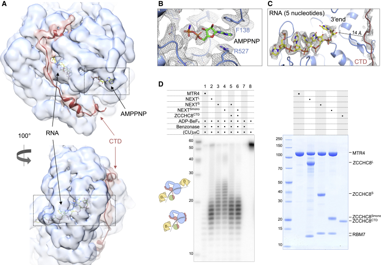
In mammalian cells, spurious transcription results in a vast repertoire of unproductive non-coding RNAs, whose deleterious accumulation is prevented by rapid decay. The nuclear exosome targeting (NEXT) complex plays a central role in directing non-functional transcripts to exosome-mediated degradation, but the structural and molecular mechanisms remain enigmatic. Here, we elucidated the architecture of the human NEXT complex, showing that it exists as a dimer of MTR4-ZCCHC8-RBM7 heterotrimers. Dimerization preconfigures the major MTR4-binding region of ZCCHC8 and arranges the two MTR4 helicases opposite to each other, with each protomer able to function on many types of RNAs. In the inactive state of the complex, the 3' end of an RNA substrate is enclosed in the MTR4 helicase channel by a ZCCHC8 C-terminal gatekeeping domain. The architecture of a NEXT-exosome assembly points to the molecular and regulatory mechanisms with which the NEXT complex guides RNA substrates to the exosome.

Keywords: NEXT; RNA degradation; RNA exosome; RNA processing; conformational regulation; cryo-EM; domain swapping; helicase; non-coding RNAs; pervasive transcription.


Conflict of interest statement

Declaration of interests E.C. is a member of the Molecular Cell advisory board.

Figures

None
Graphical abstract
Figure 1
Figure 1
Composite model of the NEXT homodimer structured core (A) Domain organization of the NEXT complex subunits MTR4, ZCCHC8, and RBM7. Domain boundaries correspond to the structural data. Dotted lines correspond to unstructured regions that were deleted in the NEXTL sample. Smaller ZCCHC8S construct used in the NEXTS reconstruction is highlighted. Abbreviations: WH, winged helix; KOW, Kyrpides-Ouzounis-Woese; NTD, N-terminal domain; CC, coiled coil; Z, Zinc finger; AIMs, arch-interacting motifs; Pro, Pro-rich domain; CTD, C-terminal domain; RRM, RNA-recognition motif. (B) Composite structural model of the NEXT homodimer obtained from interpreting cryo-EM reconstructions of the NEXTS and NEXTL samples with de novo model building and with rigid-body fitting of available crystal structures or AlphaFold predictions. Labels are indicated only for one protomer.
Figure 2
Figure 2
Interactions at the ZCCHC8 dimerization and MTR4-binding interfaces (A) Single-particle cryo-EM reconstruction of NEXTS at a global resolution of 4.5 Å, low-pass filtered to 5 Å, shown in two views, with density colored according to Figure 1B. The two protomers of ZCCHC8 and MTR4 are labeled as A and B. The box indicates the area used for the focused refinement shown in (B). (B) 4.0 Å resolution cryo-EM reconstruction after focused refinement at the dimerization module sandwiched between MTR4 KOW domains, followed by density modification (Phenix). The boxes highlight the regions in the zoom-in views of (C)–(H) where the ZCCHC8 model could be built de novo (with [C], [D], and [F] shown after an ∼90° rotation). (C) Zoom-in of the reconstruction at the dimerization module showing the density at the β-sheet with its domain-swapping topology. (D) Zoom-in of the interaction between antiparallel helices ⍺2 at the bottom of the dimerization module. (E) Zoom-in of the interaction between helices ⍺1 forming the N-terminal coiled coil. Lower map threshold was applied (PyMOL) compared with other panels, to better visualize density for the helices. (F–H) Zoom-in view at different sites of ZCCHC8-MTR4 KOW interactions: at the classical AIM (F), at the bulge (G), and at the AIM+ (H).
Figure 3
Figure 3
Monomeric form of ZCCHC8 supports MTR4-RBM7 association (A) Biochemical analysis to identify the major determinants of ZCCHC8 dimerization and MTR4-binding properties in vitro. The recombinant protein samples indicated at the top were subjected to size-exclusion chromatography experiments (analytical S200i, bottom left) and the peak fractions analyzed on a Coomassie-stained 4%–12% bis-Tris SDS-PAGE (bottom right). Deletion of the N-terminal 176 residues resulted in a monomeric NEXT mutant. (B) Biophysical analysis to quantify the contribution of ZCCHC8 dimerization on MTR4-binding. The microscale thermophoresis experiment was carried out by keeping eYFP-tagged MTR4 at a fixed concentration (50 nM) and adding increasing amounts of dimeric or monomeric ZCCHC8 and RBM7. Titrations were performed in triplicate and error bars represent standard deviation. Fitting of the experimental data curve and estimation of the dissociation constant (KD) was done with the MO software (NanoTemper technologies). (C) Cell-based analysis to assess the presence of ZCCHC8 oligomerization in vivo and its effect on MTR4-RBM7 binding. Co-immunoprecipitation experiments were carried out with exogenous MYC-tagged versions of ZCCHC8 (ev, empty vector) to assess the interaction with endogenous ZCCHC8-3F-mAID in the presence or absence of auxin (IAA) treatment. The experiment showed that full-length ZCCHC8 (residues 1–707) can interact with endogenous ZCCHC8-3F-mAID, while a mutant lacking the N-terminal 176 residues fails to dimerize but can still co-precipitate MTR4 and RBM7.
Figure 4
Figure 4
The RNA-binding domains of ZCCHC8 and RBM7 are adjacent to the MTR4 helicase domain (A) Single-particle cryo-EM reconstruction focused on a single NEXTL protomer at a global resolution of ∼7 Å. Cryo-EM density is depicted as a gray transparent surface and the composite model is shown as a ribbon. The model of the KOW domain with the ZCCHC8 AIM and AIM+ regions is from the de novo tracing from the NEXTS reconstruction (Figure 2). The model of the MTR4 DExH-box domain bound to the ZCCHC8 CTD (PDB: 6C90) (Puno and Lima, 2018) could be placed as a rigid body in the density. The model of the ZCCHC8 zinc-finger from AlphaFold and the crystal structure of the RBM7-binding module (Falk et al., 2016) (PDB: 5LXR) could be tentatively placed in the density as rigid bodies with an orientation fulfilling the connecting density features and the mass spectrometry cross-linking data (B). Other small density features at the MTR4 arch and DExH-box domains could be interpreted by predictions of the corresponding regions with AlphaFold (see also Figure S4). (B) Mass spectrometry analysis of the BS3-cross-linked monomeric mutant NEXTS (i.e., containing ZCCHC8S, residues 177–337; Figure 3) in the presence of the uridine-rich RNA and AMPPNP. Identified Lys-Lys cross-links are highlighted on the diagram and cartoon representation of the complex.
Figure 5
Figure 5
RNA is enclosed in the MTR4 helicase channel when NEXT is not active (A) Cryo-EM reconstruction from the NEXTL sample focused at the DExH-box helicase domain of MTR4. In the two views, the cryo-EM map is shown as a transparent surface at a low map threshold (Chimera) to highlight the density features at the bottom of the DExH-box domain, that could be interpreted by rigid-body fitting the corresponding crystal structure bound to the ZCCHC8 CTD (PDB: 6C90) (Puno and Lima, 2018). (B) Zoom-in view of the density of the ATP analog, AMPPNP, bound in the MTR4 ATPase site. The same cryo-EM reconstruction as above (reaching a nominal resolution of 3.4 Å) represented as mesh at a higher map threshold (PyMOL) to reveal nucleotide and amino acid details. (C) Zoom-in view of the density in the RNA-binding channel of MTR4. Meshed experimental density, at the same map threshold as in (B), surrounds 5 ribonucleotides (yellow sticks) and ZCCHC8 CTD (red ribbon). (D) RNase protection assay showing the RNA fragments obtained upon RNase treatment of the 32P body-labeled (CU)28C 57-mer RNA in the presence of the indicated protein complexes. After incubation with benzonase, the reactions’ products were analyzed by electrophoresis on a 12% acrylamide and 7 M urea gel, followed by phosphorimaging. Right side of the panel shows a Coomassie-stained 4%–12% bis-Tris SDS-PAGE with complexes used in the assay. The cartoon schematics depict how benzonase, an endonuclease, might access the RNA in our hypothesized model, resulting in a short or long footprint.
Figure 6
Figure 6
Cryo-EM reconstruction of a NEXT-exosome assembly (A) Coomassie-stained 15% SDS-PAGE gel with peak fraction from size-exclusion chromatography of a reconstituted NEXTS-exosome complex. The three NEXTS complex components are labeled in red. Two of the nuclear exosome cofactors, MPP6 and RRP6N, are fused to the EXO9 subunits RRP40 and RRP4, respectively (Gerlach et al., 2018). (B) Representative 2D classes of the dimeric NEXTS complex interacting with either one or two exosome complexes. (C) Best 3D class representing the NEXTS dimer bound to a single exosome. The cryo-EM map is shown as a transparent surface, colored according to the protein models rigid-body fitted in the density. The RBM7-binding module was fitted as in Figure 4A and the RRP6N/RRP47 module was fitted based on Gerlach et al. (2018) and Schuller et al. (2018).

Comment in

  • What's NEXT for the exosome?
    Razew M, Galej WP. Razew M, et al. Mol Cell. 2022 Jul 7;82(13):2355-2357. doi: 10.1016/j.molcel.2022.06.017. Mol Cell. 2022. PMID: 35803215

References

    1. Afonine P.V., Klaholz B.P., Moriarty N.W., Poon B.K., Sobolev O.V., Terwilliger T.C., Adams P.D., Urzhumtsev A. New tools for the analysis and validation of cryo-EM maps and atomic models. Acta Crystallogr. D Struct. Biol. 2018;74:814–840. doi: 10.1107/S2059798318009324. - DOI - PMC - PubMed
    1. Bepler T., Morin A., Rapp M., Brasch J., Shapiro L., Noble A.J., Berger B. Positive-unlabeled convolutional neural networks for particle picking in cryo-electron micrographs. Nat. Methods. 2019;16:1153–1160. doi: 10.1038/s41592-019-0575-8. - DOI - PMC - PubMed
    1. Biyani N., Righetto R.D., McLeod R., Caujolle-Bert D., Castano-Diez D., Goldie K.N., Stahlberg H. Focus: the interface between data collection and data processing in cryo-EM. J. Struct. Biol. 2017;198:124–133. doi: 10.1016/j.jsb.2017.03.007. - DOI - PubMed
    1. Bonneau F., Basquin J., Ebert J., Lorentzen E., Conti E. The yeast exosome functions as a macromolecular cage to channel RNA substrates for degradation. Cell. 2009;139:547–559. doi: 10.1016/j.cell.2009.08.042. - DOI - PubMed
    1. Butler J.S., Mitchell P. Rrp6, rrp47 and cofactors of the nuclear exosome. Adv. Exp. Med. Biol. 2010;702:91–104. doi: 10.1007/978-1-4419-7841-7_8. - DOI - PubMed

Publication types