. 2022 Jun;10(6):e004763.
doi: 10.1136/jitc-2022-004763.

Targeting of GSDMD sensitizes HCC to anti-PD-1 by activating cGAS pathway and downregulating PD-L1 expression

Affiliations

Targeting of GSDMD sensitizes HCC to anti-PD-1 by activating cGAS pathway and downregulating PD-L1 expression

Tingting Lv et al. J Immunother Cancer. 2022 Jun.

Abstract

Background: Gasdermin D (GSDMD) is well known as a downstream of inflammasomes. However, the roles of GSDMD itself in hepatocellular carcinoma (HCC) remain unclear.

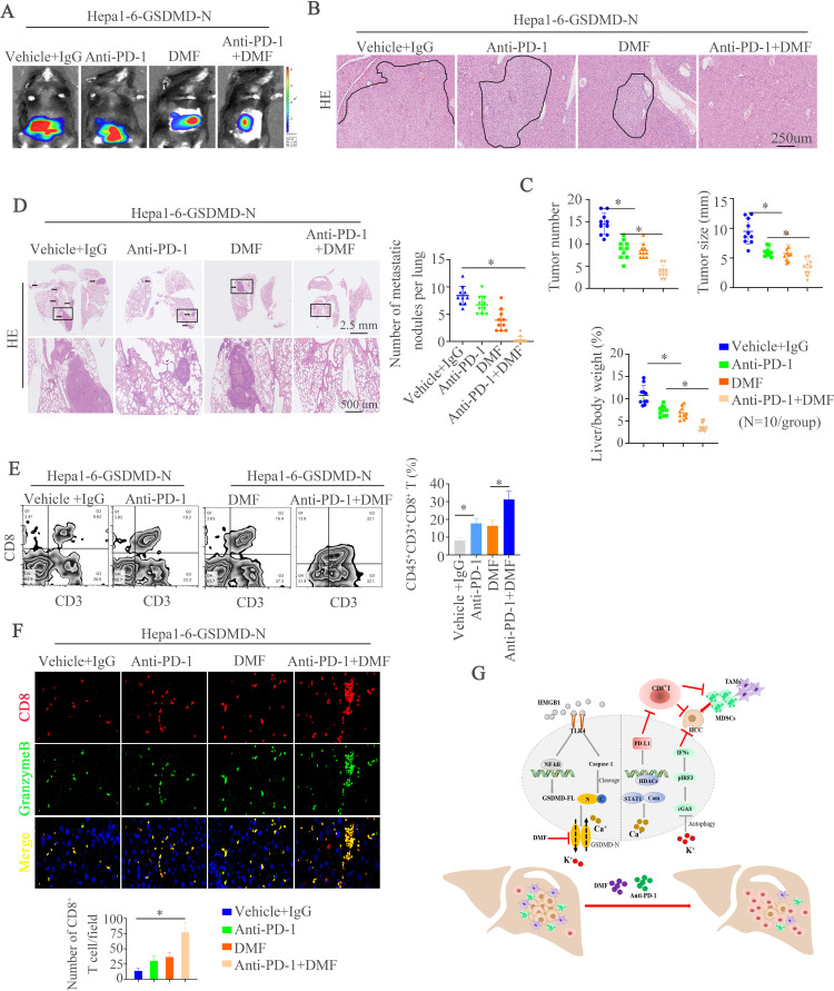
Methods: Two independent cohorts of patients with HCC were analyzed to evaluate the pathological relevance of GSDMD/pTBK1/PD-L1. GSDMD knockout (GSDMD-/-) mice, Alb-Cre mice administered with an adeno-associated virus (AAV) vector that expressed the gasdermin-N domain (AAV9-FLEX-GSDMD-N) and their wild-type littermates were used to induce hepatocarcinogenesis or metastatic HCC. Combination of anti-programmed cell death protein-1 (PD-1) and GSDMD inhibitor dimethyl fumarate (DMF) was used to test for improved therapeutic efficacy. RNA sequencing was used to explore the mechanisms how GSDMD promoted HCC progression.

Results: The expression of GSDMD and GSDMD-N was upregulated in HCC tissues or metastatic HCC tissues and positive GSDMD expression indicated grim prognosis. Diethylnitrosamine/carbon tetrachloride or thioacetamide-treated GSDMD-/- mice exhibited decreased liver tumors. In contrast, AAV9-FLEX-GSDMD-N promoted hepatocarcinogenesis. RNA sequencing manifested that knockout of GSDMD impacted the cyclic GMP-AMP synthase (cGAS) pathway and immune-associated pathway. GSDMD damped cGAS activation by promoting autophagy via outputting potassium (K+) and promoted programmed death ligand-1 (PD-L1) expression by histone deacetylases/signal transducer and activator of transcription 1 (STAT1)-induced transactivation of PD-L1 via importing calcium (Ca2+). High Mobility Group Box 1/toll-like receptor 4 (TLR4)/caspase-1 pathway contributed to the overexpression and cleavage of GSDMD. Anti-PD-1 combining with DMF largely impaired HCC progression and metastasis.

Conclusions: Targeting GSDMD could promote expression of interferons through inactivation of cGAS pathway and downregulated the PD-L1 expression. Therefore, combined anti-PD-1 and GSDMD inhibitor might serve as an effective treatment option for patients with HCC with GSDMD upregulation.

Keywords: CD8-Positive T-Lymphocytes; Immunotherapy; Inflammation; Liver Neoplasms.


Conflict of interest statement

Competing interests: None declared.

Figures

Figure 1
Figure 1
GSDMD and its cleaved GSDMD-N are upregulated in HCC tissues and indicate grim clinical prognosis. (A) RT-qPCR was used to detect GSDMD expression in normal liver tissues (n=10) and 50 paired HCC tissues. Western blotting was applied to detect expression of GSDMD-FL and GSDMD-N expression in paired HCC tissues. (B) Representative IHC images showed GSDMD expression in HCC and adjacent non-tumor tissues in two HCC cohorts. (C) Kaplan-Meier illustrated overall survival time and recurrent rate with positive or negative GSDMD expression in two independent HCC cohorts. (D) RT-qPCR was used to detect GSDMD expression in with or without recurrent patient with HCC samples (n=30) and 30 paired metastatic samples. (E) IHC showed GSDMD expression in adjacent non-tumor, HCC or metastatic tissues. (F) Western blotting exhibited GSDMD-N expression in adjacent non-tumor, HCC or metastatic tissues. GSDMD, gasdermin D; GSDMD-FL, full-length GSDMD; GSDMD-N, gasdermin-N domain; HCC, hepatocellular carcinoma; IHC, immunohistochemistry.
Figure 2
Figure 2
Deletion of GSDMD impairs hepatocarcinogenesis and overexpression of GSDMD-N promotes tumorigenesis in mice. (A) A schematic diagram for procedure of hepatocarcinogenesis induced by DEN and CCL4. (B) Representative liver and H&E images showed liver tumors of both genotypes at indicated time points. (C) Tumor numbers, maximal size and liver weight of tumors from both groups. (D) Representative IHC images illustrated proliferative hepatocytes at indicated time points measured by PCNA positive nucleus staining. (E) Representative IHC images exhibited damaged hepatocytes at indicated time points measured by γH2AX staining. (F) Masson and Sirius red staining were used to show collagenous fiber in different groups. (G) αSMA staining in liver tissues. (H) Correlation of GSDMD mRNA expression with mRNA expression of Clo1a1 and ACTA2 in non-tumor areas of hepatic tissues from WT or GSDMD-/- animals treated with DEN and CCl4. CCl4, carbon tetrachloride; DEN, diethylnitrosamine; GSDMD, gasdermin D; GSDMD-N, gasdermin-N domain; IHC, immunohistochemistry; mRNA, messenger RNA; PCNA, proliferating cell nuclear antigen; WT, wild type.
Figure 3
Figure 3
GSDMD deficiency promotes the activation of cGAS pathway in hepatocellular carcinoma. (A) The Metascape analysis stated main changed pathways between WT and GSDMD−/− mice. (B) Top 10 pathways between WT and GSDMD−/− mice were exhibited. (C) RT-qPCR analysis of indicated genes in liver tissues sorted from mice in WT and GSDMD−/− mice. (D) Protein levels of IFN-β and TNF-α was detected by ELISA in different groups. (E) Western blotting was used to analyze phosphorylated (p-)STING, STING, pTBK1, TBK1, pIRF3, IRF3, GSDMD-FL, and β-actin. (F) IF images illustrated punctate staining in different groups. (G) cGAMP amount in WT and GSDMD−/− mice was measured by UPLC/MS analysis. (H) STING monomers and dimers in lysates of WT and GSDMD−/− mice, and Huh7-control, huh7-GSDMD-N, MHCC97H-shcontrol and MHCC97H-shGSDMD stimulated with poly(dA:dT) were assessed by non-reducing polyacrylamide gel electrophoresis and western blotting. cGAS, cyclic GMP-AMP synthase; GSDMD, gasdermin D; GSDMD-FL, full-length GSDMD; IFN, interferon; TNF, tumor necrosis factor; UPLC/MS; ultra performance liquid chromatography/tandem mass spectrometry; WT, wild type.
Figure 4
Figure 4
GSDMD promotes output of intracellular K+ which regulates IFN-β expression via autophagy. (A–B) Intracellular K+ was assessed by APG4 staining in indicated groups. (C) IFN-β secretion in indicated cell stimulated with poly(dA:dT) was detected by ELISA. (D) Western blotting analyzed pSTING, STING, pTBK1, TBK1, pIRF3, IRF3, GSDMD-N, and β-actin in Huh7-GSDMD-N with or without KCl and Huh7 transfected with LV-WT or LV- I105N stimulated with poly(dA:dT). (E, F) Transmission electron microscope was shown in different groups. (G) Western blotting exhibiting LC3A/B, P62 and β-actin in different groups. (H) Western blotting exhibiting LC3A/B, P62, cGAS and β-actin in Huh7-GSDMD with or without KCl. (I) Western blotting exhibiting LC3A/B, P62, cGAS and β-actin in Huh7-GSDMD with or without Rapamycin. (J) Representative liver and H&E images illustrated liver tumors of three groups at the indicated time points. (J) cGAMP amounts in indicated group mice as measured by UPLC/MS analysis. CCl4, carbon tetrachloride; cGAS, cyclic GMP-AMP synthase; DEN, diethylnitrosamine; GSDMD, gasdermin D; GSDMD-N, gasdermin-N domain; IFN, interferon; LC3, light chain 3; UPLC/MS, ultra performance liquid chromatography/tandem mass spectrometry; WT, wild type.
Figure 5
Figure 5
GSDMD upregulates PD-L1 expression through inducing phosphorylation of STAT1. (A) Western blotting analyzed PD-L1 expression in WT and GSDMD–/– group treated with diethylnitrosamine/carbon tetrachloride or thioacetamide. (B) The mRNA expression of PD-L1 in Huh7-control, Huh7-GSDMD-N, MHCC97H-shcontrol and MHCC97H-shGSDMD. (C) The expression of PD-L1 and GSDMD-N in indicated groups was measured by western blotting. (D) CD274 promoter activity was detected after the overexpression of GSDMD-N in Huh7 cells. (E) Truncated and mutated CD274 promoter sequences and pCMV-GSDMD-N were cotransfected into Huh7 cells for testing luciferase activity. (F) CD274 promoter activity was detected after overexpression of GSDMD-N in Huh7 cells transfected with or without shSTAT1. (G) chromatin immunoprecipitation assays showed binding of STAT1 on CD274 promoter in hepatocellular carcinoma cells. (H) RT-qPCR analysis of PD-L1 mRNA after overexpression of GSDMD-N in Huhn7 cells transfected with or without shSTAT1. (I) Western blotting analyzed STAT1, pSTAT1, PD-L1, GSDMD-N and β-actin after overexpression of GSDMD-N in Huh7 cells transfected with or without shSTAT1. CCL4, carbon tetrachloride; DEN, diethylnitrosamine; GSDMD, gasdermin D; GASMD-N, gasdermin-N domain; mRNA, messenger RNA; PD-L1, programmed death ligand-1; TAA, thioacetamide.
Figure 6
Figure 6
GSDMD upregulates PD-L1 expression through Ca2+/HDACs/STAT1 pathway. (A) ChIP assays were used to illustrate binding of H4ac, H3K27ac and H3K9ac on CD274 promoter in HCC cells. (B) RT-qPCR analysis of PD-L1 mRNA in the Huh7-control, Huh7-GSDMD-N and Huh7-GSDMD-N with inhibitors of HDACs. (C) The binding of HDAC1, HDAC2 and HDAC3 on CD274 promoter in HCC cells was detected by ChIP. (D) The Ca2+ level was measured in Huh7-control and Huh7-GSDMD-N cells. (E) Immunofluorescence was used to detect calmodulin translocation in Huh7-control and Huh7-GSDMD-N cells. (F) Co-IP was used to show interact of HDAC1 and calmodulin in Huh7-control and Huh7-GSDMD-N cells. (G) Western blotting analyzed STAT1, pSTAT1, PD-L1, GSDMD-N and β-actin in indicated HCC cells. ChIP, chromatin immunoprecipitation; GSDMD, gasdermin D; GSDMD-N, gasdermin-N domain; HCC, hepatocellular carcinoma; HDAC, histone deacetylase; mRNA, messenger RNA; PD-L1, programmed death ligand-1.
Figure 7
Figure 7
HMGB1 contributes to GSDMD-FL upregulation through TLR4/ERK/P65 pathway and cleaves GSDMD-FL through activating caspase-1. (A) Huh7 cells were treated with recombinant human HMGB1 for 24 hours. GSDMD expression was detected by western blotting. (B) Truncated and mutated GSDMD promoter sequences in Huh7 cells transfected with plasmids of shcontrol or shP65 under HMGB1 treatment. (C) Western blotting analyzed P65, pP65, GSDMD-FL and β-actin in indicated HCC cells. (D) Western blotting analyzed TLR4, P65, pP65, GSDMD-FL and β-actin in indicated HCC cells. (E) ERK, JNK, p38, or PI3K pathway inhibitors were applied in Huh7 cells under HMGB1 treatment. Expression of GSDMD-FL, total and phosphorylated ERK, JNK, p38, and AKT was detected by western blotting. (F) Caspase-1 was detected in Huh7 cells treated with HMGB1 by western blotting. (G) GSDMD-N was detected in Huh7 cells treated with HMGB1 and/or Z-YVAD-FMK. GSDMD, gasdermin D; GSDMD-FL, full-length GSDMD; HCC, hepatocellular carcinoma; HMGB1, High Mobility Group Box 1; TLR4, toll-like receptor 4.
Figure 8
Figure 8
Combined GSDMD inhibitor DMF and PD-1 treatment abolishes hepatic tumorigenesis and HCC metastasis. (A) Representative bioluminescent images in mice liver. (B) Representative H&E images showed liver tumors of different groups at indicated time points. (C) Tumor numbers, maximal size and liver weight of tumors from both groups. (D) H&E staining of metastatic lung nodules was shown in different groups at indicated time points. (E) flow cytometer for CD8+ T cells in HCC tissues of HCC model after indicated treatment was displayed. (F) Immunofluorescence showed infiltration and activation of CD8+ T cells in different groups. (G) A schematic illustration elaborated the role of GSDMD on HCC metastasis. HMGB1 upregulated GSDMD expression through the ERK/P65 pathway. GSDMD overexpression inactivated cGAS pathway and induced programmed death ligand-1 expression through K+ influx and Ca2+ efflux respectively. The combined treatment of anti-PD-1 and GSDMD inhibitor DMF suppressed GSDMD-induced HCC metastasis. cGAS, cyclic GMP-AMP synthase; DMF, dimethyl fumarate; GSDMD, gasdermin D; GSDMD-N, gasdermin-N domain; HCC, hepatocellular carcinoma; HMGB1, High Mobility Group Box 1; PD-1, programmed cell death protein-1.

References

    1. Siegel RL, Miller KD, Fuchs HE, et al. . Cancer statistics, 2021. CA Cancer J Clin 2021;71:7–33. 10.3322/caac.21654 - DOI - PubMed
    1. Kelley RK, Greten TF. Hepatocellular Carcinoma - Origins and Outcomes. N Engl J Med 2021;385:280–2. 10.1056/NEJMcibr2106594 - DOI - PubMed
    1. Llovet JM, Kelley RK, Villanueva A, et al. . Hepatocellular carcinoma. Nat Rev Dis Primers 2021;7:6. 10.1038/s41572-020-00240-3 - DOI - PubMed
    1. Shi J, Gao W, Shao F. Pyroptosis: gasdermin-mediated programmed necrotic cell death. Trends Biochem Sci 2017;42:245–54. 10.1016/j.tibs.2016.10.004 - DOI - PubMed
    1. Shi J, Zhao Y, Wang K, et al. . Cleavage of GSDMD by inflammatory caspases determines pyroptotic cell death. Nature 2015;526:660–5. 10.1038/nature15514 - DOI - PubMed

Publication types

MeSH terms

Substances