. 2022 Jun;10(6):e004688.
doi: 10.1136/jitc-2022-004688.

Peripheral blood immune cell dynamics reflect antitumor immune responses and predict clinical response to immunotherapy

Affiliations

Peripheral blood immune cell dynamics reflect antitumor immune responses and predict clinical response to immunotherapy

Michael Hwang et al. J Immunother Cancer. 2022 Jun.

Abstract

Background: Despite treatment advancements with immunotherapy, our understanding of response relies on tissue-based, static tumor features such as tumor mutation burden (TMB) and programmed death-ligand 1 (PD-L1) expression. These approaches are limited in capturing the plasticity of tumor-immune system interactions under selective pressure of immune checkpoint blockade and predicting therapeutic response and long-term outcomes. Here, we investigate the relationship between serial assessment of peripheral blood cell counts and tumor burden dynamics in the context of an evolving tumor ecosystem during immune checkpoint blockade.

Methods: Using machine learning, we integrated dynamics in peripheral blood immune cell subsets, including neutrophil-lymphocyte ratio (NLR), from 239 patients with metastatic non-small cell lung cancer (NSCLC) and predicted clinical outcome with immune checkpoint blockade. We then sought to interpret NLR dynamics in the context of transcriptomic and T cell repertoire trajectories for 26 patients with early stage NSCLC who received neoadjuvant immune checkpoint blockade. We further determined the relationship between NLR dynamics, pathologic response and circulating tumor DNA (ctDNA) clearance.

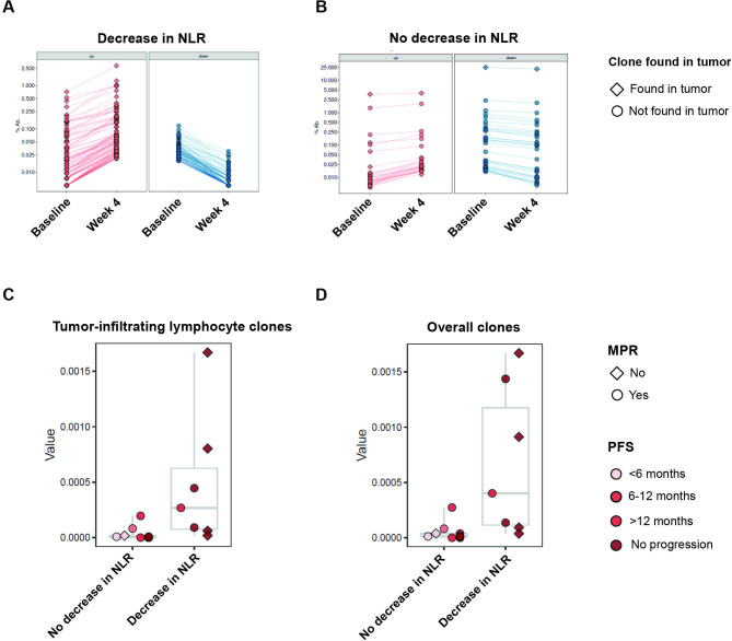
Results: Integrated dynamics of peripheral blood cell counts, predominantly NLR dynamics and changes in eosinophil levels, predicted clinical outcome, outperforming both TMB and PD-L1 expression. As early changes in NLR were a key predictor of response, we linked NLR dynamics with serial RNA sequencing deconvolution and T cell receptor sequencing to investigate differential tumor microenvironment reshaping during therapy for patients with reduction in peripheral NLR. Reductions in NLR were associated with induction of interferon-γ responses driving the expression of antigen presentation and proinflammatory gene sets coupled with reshaping of the intratumoral T cell repertoire. In addition, NLR dynamics reflected tumor regression assessed by pathological responses and complemented ctDNA kinetics in predicting long-term outcome. Elevated peripheral eosinophil levels during immune checkpoint blockade were correlated with therapeutic response in both metastatic and early stage cohorts.

Conclusions: Our findings suggest that early dynamics in peripheral blood immune cell subsets reflect changes in the tumor microenvironment and capture antitumor immune responses, ultimately reflecting clinical outcomes with immune checkpoint blockade.

Keywords: immunotherapy; translational medical research; tumor biomarkers.


Conflict of interest statement

Competing interests: VA receives research funding to her institution from Bristol-Myers Squibb and Astra Zeneca. PMF has received research funding to his institution from AstraZeneca, Bristol-Myers Squibb, Novartis, Corvus, Kyowa. He has also served as a consultant for Amgen, AstraZeneca, Bristol-Myers Squibb, Daiichi Sankyo, Iteos, Janssen, Mirati, Novartis, Sanofi and as a DSMB member for Polaris and Flame Therapeutics. KNS receives research funding to her institution from Bristol-Myers Squibb, Astra Zeneca, and Enara Bio, and holds founder’s equity in manaT Bio. VEV is a founder of Delfi Diagnostics and Personal Genome Diagnostics, serves on the Board of Directors and as a consultant for both organizations, and owns Delfi Diagnostics and Personal Genome Diagnostics stock, which are subject to certain restrictions under university policy. Additionally, Johns Hopkins University owns equity in Delfi Diagnostics and Personal Genome Diagnostics. VEV is an inventor of multiple licensed patents related to technologies from Johns Hopkins University. Some of these licenses and relationships are associated with equity or royalty payments directly to Johns Hopkins and VEV. VEV is an advisor to Bristol-Myers Squibb, Danaher, Genentech, and Takeda Pharmaceuticals. Within the last five years, VEV has been an advisor to Merck and Ignyta. These arrangements have been reviewed and approved by the Johns Hopkins University in accordance with its conflict of interest policies. JW is a consultant for Personal Genome Diagnostics, is the founder and owner of Resphera Biosciences and holds patents, royalties or other intellectual property from Personal Genomic Diagnostics. JER is in the advisory board/consultant of Oncocyte, receives speaking fees for Astrazeneca, and has received research funding to his institution from Genetech/Roche, and Verastem. JB is in the advisory board/consultant of Amgen, AstraZeneca, BMS, Genentech/Roche, Eli Lilly, GlaxoSmithKline, Merck, Sanofi and Regeneron, receives grant research funding from AstraZeneca, BMS, Genentech/Roche, Merck, RAPT Therapeutics, Inc and Revolution Medicines and is in the Data and Safety Monitoring Board/Committees of GlaxoSmithKline, Sanofi and Janssen. TS is in the advisory board/consultant of Cue Biopharma, Dracen, Innate, Nanobiotix, Merck, Sanofi, Synthekine, receives grant research funding from AstraZeneca, BMS, Cue Biopharma, Genentech/Roche, Merck, Nanobiotix, Synthekine, and is in the Data and Safety Monitoring Board/Committees of Astra Zeneca, and Nektar. VL has received research funding to his institution from AstraZeneca, Bristol-Myers Squibb, Merck, SeaGen. He has also served as a consultant for Takeda, SeaGen, Bristol-Myers Squibb, AstraZeneca, and Guardant Health.

Figures

Figure 1
Figure 1
Machine learning integration of peripheral immune cell subsets predicts durable clinical benefit to ICI. (A) Sample collection schema for 239 patients with advanced metastatic NSCLC treated with ICI-containing regimens. We employed an XGBoost machine learning approach to integrate 25 variables, including clinical characteristics and peripheral immune cell dynamics to predict clinical outcome through training an ensemble of 100 models and incorporating feature selection within 10-fold cross validation loops. (B) Receiver operator curve (ROC) for model prediction of durable clinical benefit (DCB) in the training cohort of 171 patients. (C) ROC for model prediction of DCB in an unseen testing cohort of 68 patients. (D) Shapley feature importance analysis; on the left, feature importance is listed in descending order of mean absolute SHAP value, while on the right, the directional value of features are color-coded in association with SHAP value where negative SHAP value is associated with DCB and positive SHAP values are associated with no durable benefit (NDB). AUC, area under receiver operator curve; ICIs, immune checkpoint inhibitors; NSCLC, non-small cell lung cancer; SHAP, Shapley Additive Explanations; XGBoost, eXtreme gradient boosting.
Figure 2
Figure 2
NLR dynamics point to distinct transcriptomic profiles in the tumor microenvironment. (A–C) Gene set enrichment analysis in baseline (pre-ICI treatment) tumor samples with (A) gene sets ranked by p value. Leading edge analyses are shown for (B) E2F targets and (C) MYC targets, which are enriched in patients who do not manifest a decrease in NLR. (D–F) Gene set enrichment analysis in post-ICI tumor samples with (D) gene sets ranked by p value. Leading edge analysis are shown for (E) IFN-γ and (F) antigen processing and presentation gene expression programs, which are significantly upregulated in the TME of patients with a peripheral blood decrease in NLR. (G–I) Fold change paired analysis between pre-ICI and post-ICI tumors for (G) IFN-γ, (H) antigen presentation, and (I) innate immune response gene signatures, suggesting peripheral NLR dynamics reflect an ICI-associated antitumor adaptive immune response. Dn, Down; ICI, immune checkpoint inhibition; IFN, interferon; NES, normalized enrichment score; NLR, neutrophil–lymphocyte ratio; TME, tumor microenvironment.
Figure 3
Figure 3
T cell repertoire dynamics are reflected in peripheral blood NLR dynamics. (A and B) Representative examples of TCR reshaping, signified by clonotypic expansions (red), notably of TCR clones also found in the tumor (diamond), or retractions (blue), notably of TCR clones not found in the tumor (circle), in peripheral blood, for (A) a patient with a decrease in NLR, compared with limited TCR repertoire changes in (B) a patient without a decrease in NLR, highlighting more clonotypic expansion in patients with a decrease in NLR in both overall clones and tumor-infiltrating clones. (C and D) Proportion of clonotypic expansion relative to baseline, defined as a statistically significant increase in clonotypic abundance in the on-therapy samples compared with pretreatment abundance. Compared with patients with unchanged or increased NLR, patients with decreased NLR had greater amounts of clonotypic expansion in both (C) clones identified in tumor and (D) overall clones. MPR, major pathologic response; NLR, neutrophil–lymphocyte ratio; PFS, progression-free survival; TCR, T cell receptor.
Figure 4
Figure 4
Peripheral immune cell subset dynamics are associated with response to ICI. (A) Representation of each patient in the early-stage NSCLC as a column, showing change in NLR along with clinical features, molecular and cellular features, and tumor and treatment features. Decreases in NLR and elevated on treatment eosinophil fractions were associated with major pathologic response and longer progression free survival. NLR dynamics did not appear to be associated with radiographic response, PD-L1 status, or type of immunotherapy received. Majority of patients showed stable disease by RECIST, highlighting difficulties of traditional radiography in capturing response to immunotherapy. (B) Early changes in NLR and on treatment eosinophil fraction are statistically associated with MPR, while PD-L1 and RECIST are not. χ2 test are based on two-sided testing for on treatment eosinophil fraction, RECIST, and PD-L1. Early changes in NLR was based on one-sided χ2 testing. ICI, immune checkpoint inhibitor; MPR, major pathologic response; Nivo, nivolumab; Nivo+Ipi, nivolumab+ipilimumab; NLR, neutrophil–lymphocyte ratio; NR, no response; NSCLC, non-small cell lung cancer; PD, progressive disease; PR, partial response; R, response; SD, stable disease.
Figure 5
Figure 5
NLR dynamics predict survival and complement ctDNA molecular responses. (A and B) Progression-free survival (A) and overall survival (B) stratified by NLR dynamics in the early-stage NSCLC cohort, demonstrating that a decreased NLR was significantly associated with longer PFS (log-rank p=0.0097) and OS (log-rank p=0.07). Survival curves were compared by using non-parametric log-rank test. (C–F) Comparison of ctDNA and NLR dynamics with tumor evaluation by RECIST and pathologic response at resection in representative examples. Variant allele frequency is shown on the left axis for variants confirmed to be tumor derived. Percent tumor burden and NLR value relative to baseline are shown on the right axis. For a patient with MPR (C), an early decrease in NLR captured therapeutic effect and was consistent ctDNA molecular clearance (KRAS G12C mutation) compared with RECIST tumor burden, which showed partial response. In contrast, for a patient with no tumor regression post-ICI (D), an early increase in NLR was consistent with ctDNA molecular persistence (KRAS Q61H mutation) and radiographic progressive disease. (E) In a patient where an early increase in NLR was discordant with ctDNA molecular clearance (TP53 R248L mutation), pathologic evaluation of his primary tumor and a satellite nodule revealed two separate histologies, suggesting ctDNA molecular clearance reflecting one tumor’s response and increasing NLR reflecting the other tumor’s lack of response. In a patient with undetectable ctDNA (F), early decrease in NLR accurately captured the therapeutic effect and MPR compared with RECIST tumor burden. ctDNA, circulating tumor DNA; Dec, decreased; Inc, increased; MPR, major pathologic response; NLR, neutrophil–lymphocyte ratio; NSCLC, non-small cell lung cancer; OS, overall survival; PFS, progression free survival; Unc, unchanged.

References

    1. Onoi K, Chihara Y, Uchino J, et al. . Immune checkpoint inhibitors for lung cancer treatment: a review. J Clin Med 2020;9:1362. 10.3390/jcm9051362 - DOI - PMC - PubMed
    1. Assi HI, Kamphorst AO, Moukalled NM, et al. . Immune checkpoint inhibitors in advanced non-small cell lung cancer. Cancer 2018;124:248–61. 10.1002/cncr.31105 - DOI - PubMed
    1. Hellmann MD, Paz-Ares L, Bernabe Caro R, et al. . Nivolumab plus ipilimumab in advanced non-small-cell lung cancer. N Engl J Med 2019;381:2020–31. 10.1056/NEJMoa1910231 - DOI - PubMed
    1. Doroshow DB, Sanmamed MF, Hastings K, et al. . Immunotherapy in non-small cell lung cancer: facts and hopes. Clin Cancer Res 2019;25:4592–602. 10.1158/1078-0432.CCR-18-1538 - DOI - PMC - PubMed
    1. Anagnostou V, Niknafs N, Marrone K, et al. . Multimodal genomic features predict outcome of immune checkpoint blockade in non-small-cell lung cancer. Nat Cancer 2020;1:99–111. 10.1038/s43018-019-0008-8 - DOI - PMC - PubMed

Publication types