

Extramedullary haematopoiesis presenting as an adnexal mass in a patient with β-thalassaemia
- PMID: 35688572
- PMCID: PMC9189770
- DOI: 10.1136/bcr-2022-249422
Extramedullary haematopoiesis presenting as an adnexal mass in a patient with β-thalassaemia
Abstract
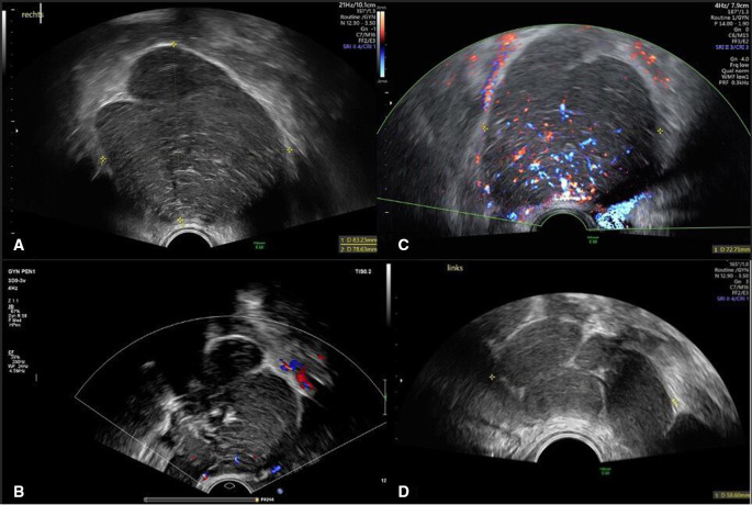
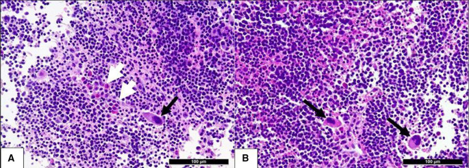
Solid masses of the ovaries raise the suspicion of malignancy or metastasis and require histological diagnosis. Extramedullary haematopoesis (EMH) is a rare histological finding of a mass of the adnexa. The sonographic pattern of EMH has rarely been described in the literature. Transvaginal biopsy of EMH has not been reported in the literature. We present a case of adnexal EMH in a patient affected with β-thalassaemia, and we performed a narrative review. Only in our case, the sonographic pattern was described, and a transvaginal ultrasound-guided core biopsy was used. Assessing patients' medical history and correlating it to the findings of diagnostic imaging is of paramount importance when evaluating patients with adnexal masses. The correct interpretation of sonographic images can avoid unnecessarily invasive procedures. A transvaginal biopsy could be a safe, easy and well-tolerated method to gain definite histological diagnosis in cases where a primary ovarian malignancy is not suspected.
Keywords: Haematology (drugs and medicines); Obstetrics, gynaecology and fertility; Radiology; Ultrasonography.
© BMJ Publishing Group Limited 2022. No commercial re-use. See rights and permissions. Published by BMJ.
Conflict of interest statement
Competing interests: None declared.
Figures
References
-
- Huang C-W, Hsueh S, Chang M-Y. Agnogenic myeloid metaplasia in an ovarian steroid cell tumor with virilization: a case report. J Reprod Med 2004;49:765–8. - PubMed
-
- Eapen SS, Narayan R, Khan A. A case of unusual extra-medullary hematopoiesis. Journal of Clinical Oncology 2004;22:6698. 10.1200/jco.2004.22.90140.6698 - DOI
-
- Timmerman D, Valentin L, Bourne TH, et al. . Terms, definitions and measurements to describe the sonographic features of adnexal tumors: a consensus opinion from the International ovarian tumor analysis (iota) group. Ultrasound Obstet Gynecol 2000;16:500–5. 10.1046/j.1469-0705.2000.00287.x - DOI - PubMed
Publication types
MeSH terms
LinkOut - more resources
Full Text Sources
Medical