. 2022 Jun;27(6):066001.
doi: 10.1117/1.JBO.27.6.066001.

Mouthwash as a non-invasive method of indocyanine green delivery for near-infrared fluorescence dental imaging

Affiliations

Mouthwash as a non-invasive method of indocyanine green delivery for near-infrared fluorescence dental imaging

Zhongqiang Li et al. J Biomed Opt. 2022 Jun.

Abstract

Significance: X-ray imaging serves as the mainstream imaging in dentistry, but it involves risk of ionizing radiation.

Aim: This study presents the feasibility of indocyanine green-assisted near-infrared fluorescence (ICG-NIRF) dental imaging with 785-nm NIR laser in the first (ICG-NIRF-I: 700 to 1000 nm) and second (ICG-NIRF-II: 1000 to 1700 nm) NIR wavelengths.

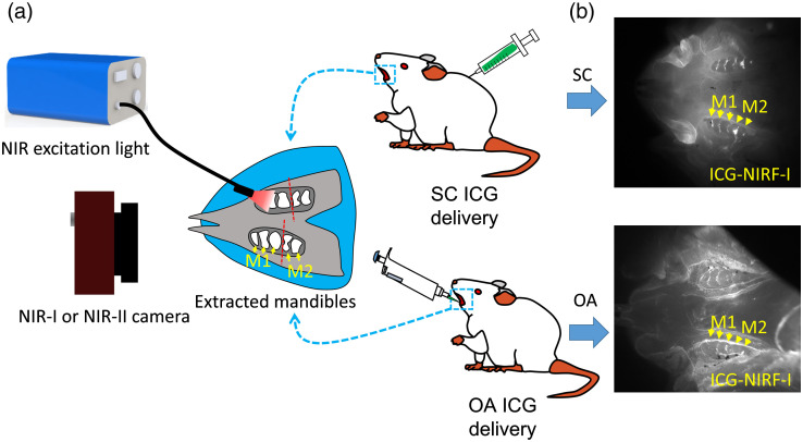
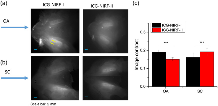
Approach: Sprague Dawley rats with different postnatal days were used as animal models. ICG, as a fluorescence agent, was delivered to dental structures by subcutaneous injection (SC) and oral administration (OA).

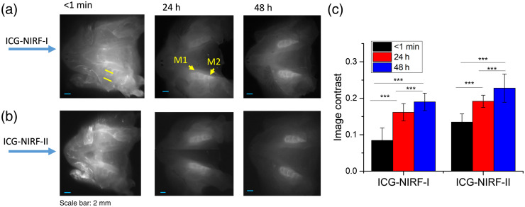
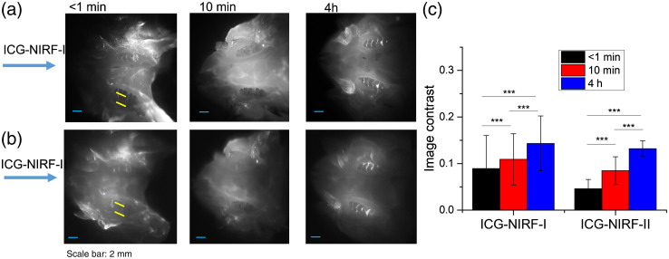
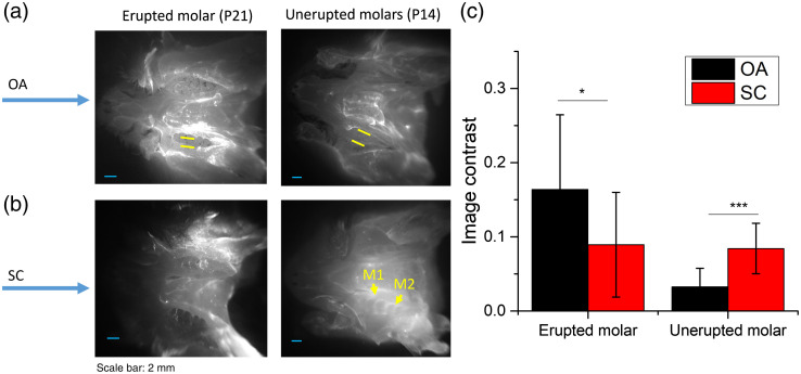
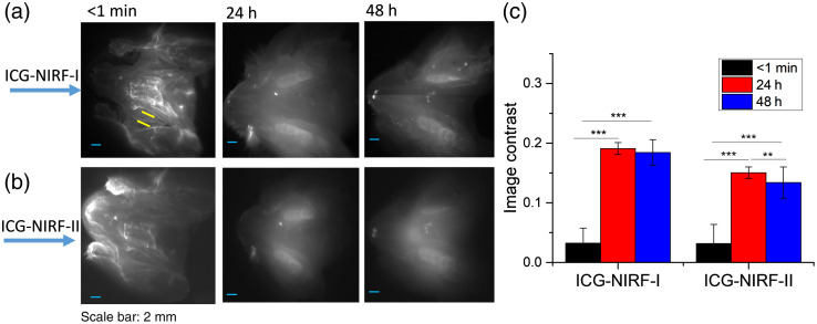
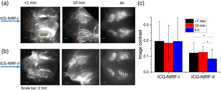
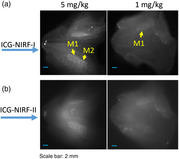
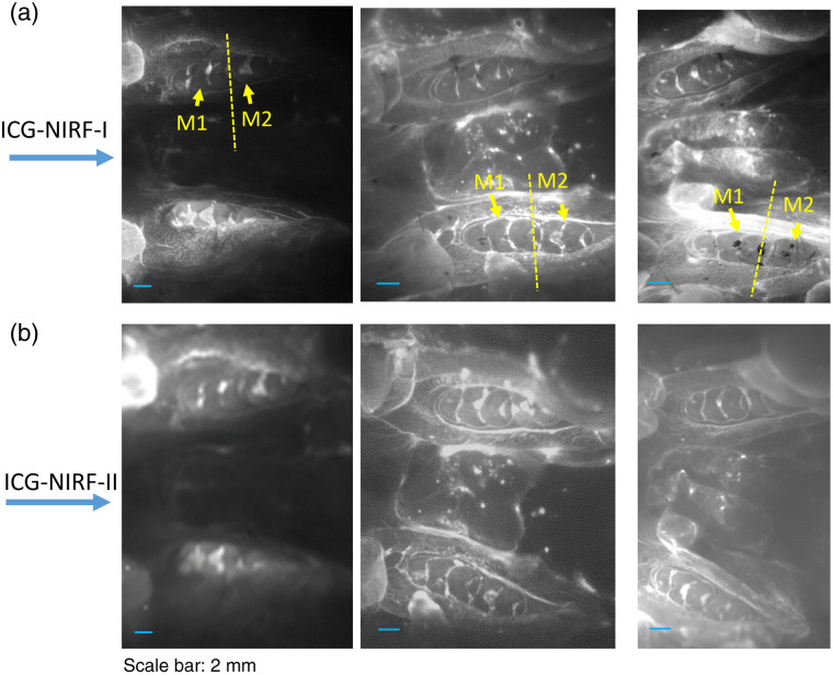
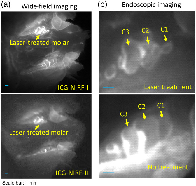
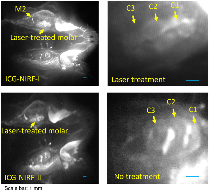
Results: For SC method, erupted and unerupted molars could be observed from ICG-NIRF images at a short imaging time (<1 min). ICG-NIRF-II could achieve a better image contrast in unerupted molars at 24 h after ICG injection. The OA could serve as a non-invasive method for ICG delivery; it could also cause the glow-in-dark effect in unerupted molars. For erupted molars, OA can be considered as mouthwash and exhibits outstanding performance for delivery of ICG dye; erupted molar structures could be observed at a short imaging time (<1 min) and low ICG dose (0.05 mg / kg).

Conclusions: Overall, ICG-NIRF with mouthwash could perform in-vivo dental imaging in two NIR wavelengths at a short time and low ICG dose.

Keywords: indocyanine green; indocyanine green delivery; mouthwash; near-infrared fluorescence dental imaging; oral administration.


Figures

Fig. 1
Fig. 1
Schematic diagram of ICG-NIRF dental imaging with SC and OA ICG delivery. (a) Schematic diagram and (b) typical ICG-NIRF images of P21 erupted molars with SC (4 h) and OA (10 min) methods. M1: first molar; M2: second molar.
Fig. 2
Fig. 2
The SC-based ICG-NIRF imaging for unerupted molars under the first (ICG-NIRF-I) and second (ICG-NIRF-II) NIR windows. (a) ICG-NIRF-I; (b) ICG-NIRF-II; and (c) image contrast of ICG-NIRF-I and ICG-NIRF-II under different imaging times. M1: the first molar; M2: the second molar. ICG dose: 5  mg/kg. * is significant at <0.05; ** is significant at <0.01; and *** is significant at <0.001.
Fig. 3
Fig. 3
The SI-based ICG-NIRF dental imaging of the erupted molars (P21) under NIRF-I and NIRF-II. (a) ICG-NIRF-I; (b) ICG-NIRF-II; and (c) image contrast of ICG-NIRF-I and ICG-NIRF-II under different imaging times. ICG dose: 5  mg/kg. * is significant at <0.05; ** is significant at <0.01; and *** is significant at <0.001.
Fig. 4
Fig. 4
Comparison of OA and SC ICG delivery, erupted and unerupted molars were imaged at short imaging time (<1  min) with ICG-NIRF-I imaging. (a) Erupted and unerupted molars by using OA ICG delivery. (b) Erupted and unerupted molars by using SC ICG delivery; (c) Image contrast of OA and SC method. M1: the first molar; M2: the second molar. ICG dose: 5  mg/kg. * is significant at <0.05; ** is significant at <0.01; and *** is significant at <0.001.
Fig. 5
Fig. 5
Comparison of OA-based and SC-based ICG-NIRF dental imaging in the unerupted molars at long imaging time (24 h). (a) The ICG-NIRF dental imaging with OA ICG delivery; (b) ICG-NIRF dental imaging with SC ICG delivery; and (c) image contrast of OA and SC methods. M1: the first molar; M2: the second molar. ICG dose: 5  mg/kg. * is significant at <0.05; ** is significant at <0.01; and *** is significant at <0.001.
Fig. 6
Fig. 6
The OA-based ICG-NIRF imaging for unerupted molars under the first (ICG-NIRF-I) and second (ICG-NIRF-II) NIR windows. (a) ICG-NIRF-I; (b) ICG-NIRF-II; and (c) image contrast of ICG-NIRF-I and ICG-NIRF-II under different imaging times. * is significant at <0.05; ** is significant at <0.01; and *** is significant at <0.001.
Fig. 7
Fig. 7
The OA-based ICG-NIRF dental imaging for the erupted molars (P21) under NIRF-I and NIRF-II. (a) ICG-NIRF-I; (b) ICG-NIRF-II; and (c) image contrast of ICG-NIRF-I and ICG-NIRF-II under different imaging times. * is significant at <0.05; ** is significant at <0.01; and *** is significant at <0.001.
Fig. 8
Fig. 8
The OA-based ICG-NIRF dental imaging of the unerupted molars (P14) with low (1  mg/kg) and high (5  mg/kg) ICG doses, and imaging after 48 h of the OA ICG delivery. (a) ICG-NIRF-I dental imaging with low and high ICG and (b) ICG-NIRF-II dental imaging with low and high ICG.
Fig. 9
Fig. 9
The OA-based ICG-NIRF dental imaging of the erupted molars (P60 young adult rats) with ICG dose ranging from 0.05 to 1  mg/kg, and imaging under NIRF-I and NIRF-II. (a) ICG-NIRF-I dental imaging and (b) ICG-NIRF-II dental imaging.
Fig. 10
Fig. 10
The OA-based ICG-NIRF imaging for the laser-treated abnormal erupted molars in P21 rats. (a) Wide-field pictures of abnormal erupted molar. (b) The endoscopic ICG-NIRF images of abnormal erupted molar and normal molar. C1: the first cusps; C2: the second cusps; and C2: the third cusps.
Fig. 11
Fig. 11
The OA-based ICG-NIRF imaging for the laser-treated abnormal erupted molars in young adult (P60) rats. (a) Wide-field pictures of abnormal erupted molar. (b) The endoscopic ICG-NIRF images of abnormal erupted molar and normal molar. C1: the first cusp; C2: the second cusp; and C2: the third cusp.

Similar articles

Cited by

References

    1. Vandenberghe B., Jacobs R., Bosmans H., “Modern dental imaging: a review of the current technology and clinical applications in dental practice,” Eur. Radiol. 20(11), 2637–2655 (2010).10.1007/s00330-010-1836-1 - DOI - PubMed
    1. A. D. Association, Dental Radiographic Examinations: Recommendations for Patient Selection and Limiting Radiation Exposure, ADA, Chicago: (2012).
    1. Kiljunen T., et al. , “Dental cone beam CT: a review,” Phys. Med. 31(8), 844–860 (2015).10.1016/j.ejmp.2015.09.004 - DOI - PubMed
    1. Shah N., Bansal N., Logani A., “Recent advances in imaging technologies in dentistry,” World J. Radiol. 6(10), 794–807 (2014).10.4329/wjr.v6.i10.794 - DOI - PMC - PubMed
    1. Tymofiyeva O., et al. , “Three-dimensional localization of impacted teeth using magnetic resonance imaging,” Clin. Oral. Investig. 14(2), 169–176 (2010).10.1007/s00784-009-0277-1 - DOI - PubMed

Publication types