. 2022 Jul 19;121(14):2767-2780.
doi: 10.1016/j.bpj.2022.06.012. Epub 2022 Jun 9.

Heme-bound tyrosine vibrations in hemoglobin M: Resonance Raman, crystallography, and DFT calculation

Affiliations

Heme-bound tyrosine vibrations in hemoglobin M: Resonance Raman, crystallography, and DFT calculation

Shigenori Nagatomo et al. Biophys J. .

Abstract

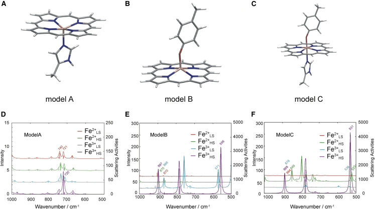
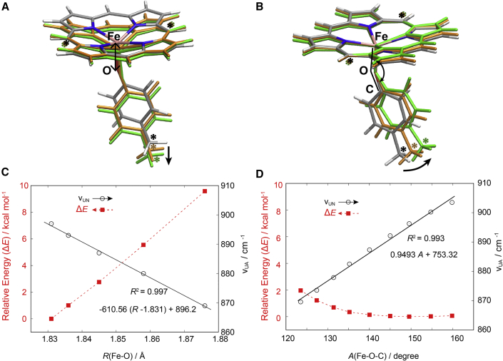
Hemoglobins M (Hbs M) are human hemoglobin variants in which either the α or β subunit contains a ferric heme in the α2β2 tetramer. Though the ferric subunit cannot bind O2, it regulates O2 affinity of its counterpart ferrous subunit. We have investigated resonance Raman spectra of two Hbs, M Iwate (α87His → tyrosine [Tyr]) and M Boston (α58His → Tyr), having tyrosine as a heme axial ligand at proximal and distal positions, respectively, that exhibit unassigned resonance Raman bands arising from ferric (not ferrous) hemes at 899 and 876 cm-1. Our quantum chemical calculations using density functional theory on Fe-porphyrin models with p-cresol and/or 4-methylimidazole showed that the unassigned bands correspond to the breathing-like modes of Fe3+-bound Tyr and are sensitive to the Fe-O-C(Tyr) angle. Based on the frequencies of the Raman bands, the Fe-O-C(Tyr) angles of Hbs M Iwate and M Boston were predicted to be 153.5° and 129.2°, respectively. Consistent with this prediction, x-ray crystallographic analysis showed that the Fe-O-C(Tyr) angles of Hbs M Iwate and M Boston in the T quaternary structure were 153.6° and 134.6°, respectively. It also showed a similar Fe-O bond length (1.96 and 1.97 Å) and different tilting angles.


Conflict of interest statement

Declaration of interests The authors declare no competing interests.

Figures

None
Graphical abstract
Figure 1
Figure 1
Oxidation and coordination state of heme in Hb M Iwate, Hb M Boston, and Hb A.
Figure 2
Figure 2
Resonance Raman spectra of Hb M Iwate of oxy forms excited at 488 nm. We used acetate buffer (pH 4.9), phosphate buffer (from pH 5.5 to 7.7), and borate buffer (pH 9.3). Sodium sulfate was 50 mM. The band of SO42- at 981 cm-1 serves as an intensity standard. Bands marked by asterisk () denote bands arising from cell glass containing sample. The bands marked by Y# indicate an internal vibration of Tyr, and those marked by ν# do ring (in-plane) vibrations of porphyrin, for both of which # denotes a mode number (7,34,35). To see this figure in color, go online.
Figure 3
Figure 3
Resonance Raman spectra of Hb M Iwate (black) and Hb M Boston (brown) of deoxy forms excited at 488 nm. We used phosphate buffer. To see this figure in color, go online.
Figure 4
Figure 4
Resonance Raman spectra of Hb M Iwate and Hb M Boston of deoxy forms (αFe3+βFe2+-deoxy) excited at 442 nm. We used phosphate buffer. To see this figure in color, go online.
Figure 5
Figure 5
(A–C) Optimized structures of the Fe-porphyrin models: (A) porphyrin and 4-methylimidazole (4-MeIm), (B) porphyrin and p-Cre, and (C) porphyrin, 4-MeIm and p-Cre. (D–F) Raman spectra calculated by the UB3LYP/VDZP theoretical level for the models (A)–(C). To see this figure in color, go online.
Figure 6
Figure 6
(A and B) The theoretical model (B in Fig. 5) used to evaluate the dependences of the Fe-O bond length (R(Fe-O)) and Fe-O-C angle (A(Fe-O-C)) on the unassigned Raman mode. Vibrational frequencies were calculated under fixing three C atoms marked with an asterisk () in the porphyrin methine and p-cresol methyl group. (C and D) The calculated vibrational frequencies and the relative energies plotted in black and red colors against the Fe-O bond length and Fe-O-C angle. The vibrational frequencies are fitted well on the least square lines. To see this figure in color, go online.
Figure 7
Figure 7
The calculated vibrational frequency of the Fe-O mode around 556 cm-1 plotted in black and red colors against the Fe-O bond length (A) and Fe-O-C angle (B). The vibrational frequencies are fitted well on the least square lines. To see this figure in color, go online.
Figure 8
Figure 8
(a and b) The |2mFo-DFc| electron density map around the T-state α1 heme in Hb M Iwate (a) and Hb M Boston (b), contoured at the level of 1.2 σ. Each structure is shown as a stick-and-sphere model colored by atom type: oxygen, red; nitrogen, blue; carbon, green; Fe, red sphere. Figures were produced using PyMOL. The electron density maps for the other subunits are similar (Fig. S3). To see this figure in color, go online.
Figure 9
Figure 9
Vibrational modes of the 550 and 900 cm-1 at different Fe-O-C angles (A(Fe-O-C) = 170° and 140°). To see this figure in color, go online.

References

    1. Perutz M.F. Regulation of oxygen affinity of hemoglobin: influence of structure of the globin on the heme iron. Annu. Rev. Biochem. 1979;48:327–386. doi: 10.1146/annurev.bi.48.070179.001551. - DOI - PubMed
    1. Imai K. Cambridge University Press; 1982. Allosteric Effects in Haemoglobin.
    1. Hayashi A., Suzuki T., et al. Watari H. Changes in EPR spectra of M-type abnormal haemoglobins induced by deoxygenation and their implication for the haem-haem interaction. Biochim. Biophys. Acta. 1967;147:407–409. doi: 10.1016/0005-2795(67)90426-6. - DOI - PubMed
    1. Nishikura K., Sugita Y., et al. Yoneyama Y. Ethylisocyanide equilibria of hemoglobins M Iwate, M Boston, M Hyde Park, M Saskatoon, and M Milwaukee-I in half-ferric and fully reduced states. J. Biol. Chem. 1975;250:6679–6685. doi: 10.1016/s0021-9258(19)40987-3. - DOI - PubMed
    1. Fung L.W.-M., Minton A.P., et al. Ho C. Proton nuclear magnetic resonance studies of hemoglobin M Milwaukee and their implications concerning the mechanism of cooperative oxygenation of hemoglobin. Biochemistry. 1977;16:1452–1462. doi: 10.1021/bi00626a033. - DOI - PubMed

Publication types

LinkOut - more resources