Review
. 2023 May 2;119(3):614-630.
doi: 10.1093/cvr/cvac093.

Adiposity-associated atrial fibrillation: molecular determinants, mechanisms, and clinical significance

Affiliations
Review

Adiposity-associated atrial fibrillation: molecular determinants, mechanisms, and clinical significance

Monika Gawałko et al. Cardiovasc Res. .

Abstract

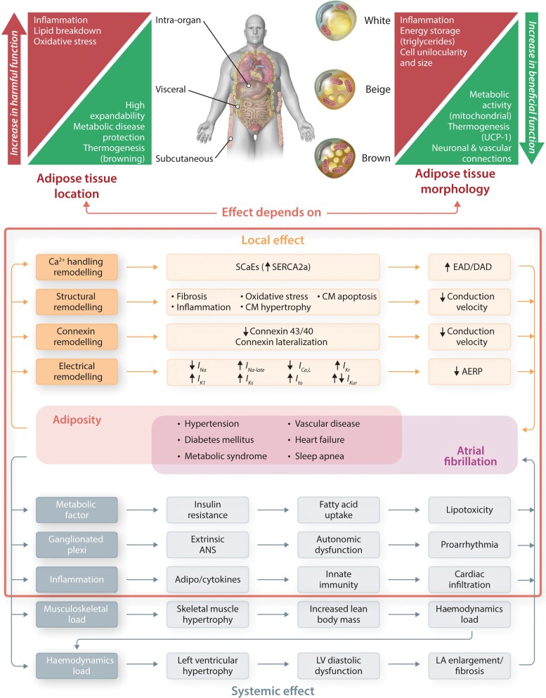
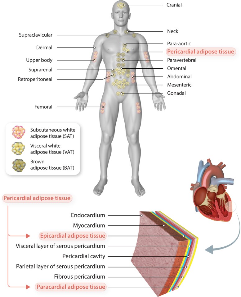
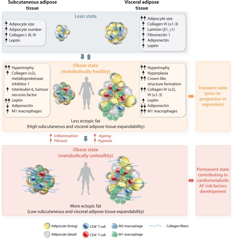
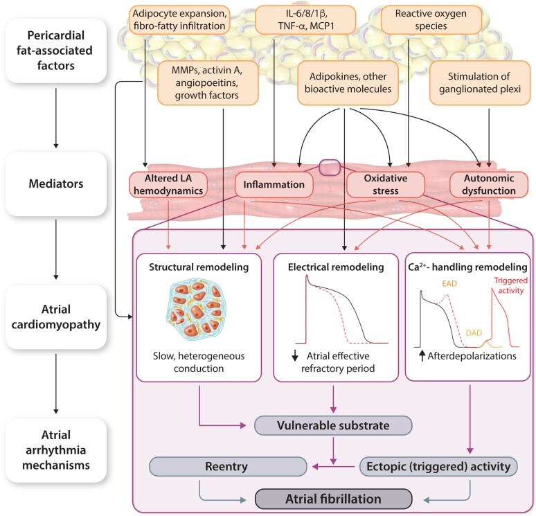
Obesity is an important contributing factor to the pathophysiology of atrial fibrillation (AF) and its complications by causing systemic changes, such as altered haemodynamic, increased sympathetic tone, and low-grade chronic inflammatory state. In addition, adipose tissue is a metabolically active organ that comprises various types of fat deposits with discrete composition and localization that show distinct functions. Fatty tissue differentially affects the evolution of AF, with highly secretory active visceral fat surrounding the heart generally having a more potent influence than the rather inert subcutaneous fat. A variety of proinflammatory, profibrotic, and vasoconstrictive mediators are secreted by adipose tissue, particularly originating from cardiac fat, that promote atrial remodelling and increase the susceptibility to AF. In this review, we address the role of obesity-related factors and in particular specific adipose tissue depots in driving AF risk. We discuss the distinct effects of key secreted adipokines from different adipose tissue depots and their participation in cardiac remodelling. The possible mechanistic basis and molecular determinants of adiposity-related AF are discussed, and finally, we highlight important gaps in current knowledge, areas requiring future investigation, and implications for clinical management.

Keywords: Adipokines; Atrial fibrillation; Epicardial adipose tissue; NLRP3 inflammasome; Obesity; Subcutaneous adipose tissue; Visceral adipose tissue.


Conflict of interest statement

Conflict of interest: A.S., N.L., I.A.-T., T.J., D.L., S.N., J.H., and A.F.: none. M.G. received grant from Pfizer and Specialized Research Fellowship (Club 30 of PCS) grant. D.D. is a member of the Scientific Advisory Boards of Omeicos Therapeutics GmbH and Acesion Pharma.

Figures

Graphical abstract
Graphical abstract
Cellular, localization, and functional heterogeneity of adipose tissue. CM, cardiomyocyte; see Tables 1 and 2. Visceral and subcutaneous adipose tissue are two main human fat depots that represent different metabolically and proinflammatory profile (upper left panel). Adipocytes are divided into different cell types with distinct morphological and functional characteristics, including white, beige, and brown adipocytes (upper right panel). Pink adipocytes are not discussed in this review, since they mainly occur in pregnancy. Adiposity together with accompanying obesity-related comorbidities (metabolic syndrome, diabetes, hypertension, heart failure, sleep apnoea) promote atrial fibrillation through systemic effects (bottom left) via haemodynamic, metabolic, neurohormonal, and proinflammatory factors, and through local effects promoting a proarrhythmic atrial cardiomyopathy involving Ca2+ handling, structural, connexin, and electrical remodelling (bottom right). Depending on location and morphology, adipose tissue might contribute to proarrhythmia by causing local and systemic effects (red frame).
Figure 1
Figure 1
Types and localization of adipose tissue depots. In humans, BAT is localized mainly around the shoulders and ribs. Visceral WAT surrounds intra-abdominal organs, whereas subcutaneous WAT spreads throughout the body beneath the skin. Pericardial adipose tissue, a subtype of VAT, comprises both the epicardial and paracardial adipose tissue layers. Epicardial fat lies between the visceral pericardium and myocardium, whereas paracardial fat is located external to the fibrous pericardium.
Figure 2
Figure 2
Schematic representation of the cellular dynamics of adipose tissue depots associated with obesity. In lean state (grey), VAT is characterized by large adipocytes, whereas SAT is characterized by a larger number of adipocytes, with VAT and SAT differing in their content of fibrotic factors and adipokines. During obesity, VAT is characterized by hyperplasia of adipocyte precursors and hypertrophy of mature adipocytes, while SAT adipocytes preferentially undergo hypertrophy. Weight gain triggers cell growth of VAT (vs. SAT) adipocytes, along with a reduction in adiponectin expression in VAT. This state is typical for ‘metabolically healthy’ individuals with high expandability of SAT and VAT, resulting in a low degree of ectopic fat storage (yellow). This state can regress or progress. As obesity progresses, extracellular matrix rigidity, composition, and remodelling alter adipose tissue expandability by physically limiting adipocyte hypertrophy and hyperplasia. Once the capacity to store lipids is reached, lipids begin to accumulate at ectopic sites. This process is modulated by genetic and environmental factors, and for VAT is further restricted by the available space in the abdominal cavity. This obese state describes ‘metabolically unhealthy’ individuals with low expandability of SAT and VAT resulting in a high rate of ectopic fat storage (red) that promotes insulin resistance, apoptosis and inflammation in places of residence (heart, liver, pancreas), thereby contributing to the development of cardiometabolic risk factors for AF.
Figure 3
Figure 3
Role of pericardial fat in AF promotion. DAD, delayed afterdepolarization; EAD, early afterdepolarization; see Table 1. Pericardial fat-associated factors (adipocyte expansion/infiltration, inflammatory signalling molecules, growth factors, adipokines, reactive oxygen species, and stimulation of ganglionated plexi) stimulate the development of an atrial cardiomyopathy via a wide range of mediators (inflammation, oxidative stress, mechanical, and autonomic dysfunction). The obesity-induced atrial cardiomyopathy includes a vulnerable substrate, consisting of re-entry-promoting structural, connexin, and electrical remodelling, as well as Ca2+-handling remodelling leading to triggered activity via early and delayed afterdepolarizations. Together, these arrhythmia mechanisms promote the initiation and maintenance of AF. IL, interleukin; MCP1, monocyte chemoattractant protein-1; TNF-α, tumour necrosis factor alpha.

References

    1. Maffetone PB, Rivera-Dominguez I, Laursen PB. Overfat and underfat: new terms and definitions long overdue. Front Public Health 2016;4:279. - PMC - PubMed
    1. Powell-Wiley TM, Poirier P, Burke LE, Despres JP, Gordon-Larsen P, Lavie CJ, Lear SA, Ndumele CE, Neeland IJ, Sanders P, St-Onge MP; American Heart Association Council on Lifestyle and Cardiometabolic Health; Council on Cardiovascular and Stroke Nursing; Council on Clinical Cardiology; Council on Epidemiology and Prevention; and Stroke Council . Obesity and cardiovascular disease: a scientific statement from the American Heart Association. Circulation 2021;143:e984–e1010. - PMC - PubMed
    1. Schoettl T, Fischer IP, Ussar S. Heterogeneity of adipose tissue in development and metabolic function. J Exp Biol 2018;221:jeb162958. - PubMed
    1. Russo L, Lumeng CN. Properties and functions of adipose tissue macrophages in obesity. Immunology 2018;155:407–417. - PMC - PubMed
    1. Omran F, Christian M. Inflammatory signaling and brown fat activity. Front Endocrinol(Lausanne) 2020;11:156. - PMC - PubMed

Publication types