. 2022 Jul 25;14(7):mfac041.
doi: 10.1093/mtomcs/mfac041.

A ferrocene-containing nucleoside analogue targets DNA replication in pancreatic cancer cells

Affiliations

A ferrocene-containing nucleoside analogue targets DNA replication in pancreatic cancer cells

Marium Rana et al. Metallomics. .

Abstract

Pancreatic ductal adenocarcinoma (PDAC) is a disease that remains refractory to existing treatments including the nucleoside analogue gemcitabine. In the current study we demonstrate that an organometallic nucleoside analogue, the ferronucleoside 1-(S,Rp), is cytotoxic in a panel of PDAC cell lines including gemcitabine-resistant MIAPaCa2, with IC50 values comparable to cisplatin. Biochemical studies show that the mechanism of action is inhibition of DNA replication, S-phase cell cycle arrest and stalling of DNA-replication forks, which were directly observed at single molecule resolution by DNA-fibre fluorography. In agreement with this, transcriptional changes following treatment with 1-(S,Rp) include activation of three of the four genes (HUS1, RAD1, RAD17) of the 9-1-1 check point complex clamp and two of the three genes (MRE11, NBN) that form the MRN complex as well as activation of multiple downstream targets. Furthermore, there was evidence of phosphorylation of checkpoint kinases 1 and 2 as well as RPA1 and gamma H2AX, all of which are considered biochemical markers of replication stress. Studies in p53-deficient cell lines showed activation of CDKN1A (p21) and GADD45A by 1-(S,Rp) was at least partially independent of p53. In conclusion, because of its potency and activity in gemcitabine-resistant cells, 1-(S,Rp) is a promising candidate molecule for development of new treatments for PDAC.

Keywords: DNA replication; Ferrocene; nucleoside analogue; pancreatic cancer; replication fork arrest.


Conflict of interest statement

The authors declare no conflicts of interest.

Figures

Graphical Abstract
Graphical Abstract
1-(S,Rp) stalls DNA replication forks resulting in S-phase cell cycle arrest.
Fig. 1
Fig. 1
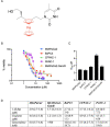
Compound 1-(S,Rp) whose chemical structure is shown in (A) with ferrocene highlighted in red is cytotoxic to a panel of pancreatic ductal adenoma carcinoma cells. (B) Cytotoxicity curves in pancreatic ductal adenoma carcinoma (PDAC) cell lines treated with 1-(S,Rp) for 72 h as assessed by the MTT [3-(4,5-dimethylthiazol-2-yl)-2,5-diphenyltetrazolium bromide] assay. The results represent the mean of three independent biological experiments (n = 3). (C) Modelled IC50 (±SD) values of 1-(S,Rp) (variable slope 4 parameter). (D) Comparison of 72 h IC50 values for 1-(S,Rp), cisplatin expressed in micromoles and gemcitabine HCl, expressed in nanomoles in the panel of five pancreatic ductal adenoma cells. The values in parentheses are the 95% CIs. All results represent the mean from three independent biological experiments (n = 3) and are normalized to vehicle control.
Fig. 2
Fig. 2
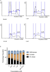
Compound 1-(S,Rp) causes concentration-dependent S-phase arrest in MIAPaCa2 pancreatic ductal adenoma carcinoma cells. Cells were treated with 1-(S,Rp) (0–5 μM) for 24 h before staining with propidium iodide and analysis by flow cytometry. (A) Representative histograms from cells treated with 0, 1, 2.5 and 5 μM 1-(S,Rp). (B) Graphical representation of data, the results represent the mean of three independent biological experiments (±SD, n = 3). *, ** and *** statistically significantly different as assessed by 2-way ANOVA followed by Dunnett's multiple comparison t-test. As a positive control, cells were also treated with 0.5 μM gemcitabine.
Fig. 3
Fig. 3
Compound 1-(S,Rp) inhibits replication of genomic DNA in MIAPaCa2 pancreatic ductal adenoma carcinoma cells. (A) Incorporation of EdU into replicating DNA as assessed by flow cytometry following treatment of cells with 1-(S,Rp) for 24 h. *** Significantly different from untreated control (1-way ANOVA followed by a post hoc Dunnett's t-test). (B) Percentage of EdU incorporating and non-incorporating cells following treatment with 1-(S,Rp) (24 h) as quantified using the criteria outlined in (C) for a representative experiment. (D) Confocal microscopy confirms reduced EdU incorporation (green fluorescence) into genomic DNA in the nuclei of cells counter stained with DAPI (blue fluorescence). (E) Median cellular labelling with CellTrace Far Red. (F) Representative histograms from an individual experiment. The results represent the mean of three independent biological experiments (±SD, n = 3). *** Statistically significant from control (P < 0.001, 1-way ANOVA followed by a post hoc Dunnett's t-test). The calculated EC50 of 1-(S,Rp) was 1.3 μM (95% CI 0.9–1.9 μM).
Fig. 4
Fig. 4
List of 39 genes related to DNA repair that are statistically significantly upregulated in MIAPaCa2 cells following treatment with 1-(S,Rp) (10 μM, 24 h). (A) Graphed as fold change (2−ddCq) and (B) as a heat map for clarity. The results represent the mean of three independent biological experiments (±SD, n = 3). Statistically significantly changed genes plotted relative to GAPDH as 2−dCq are shown in Supplementary Fig. S3.
Fig. 5
Fig. 5
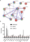
Compound 1-(S,Rp) induces a conserved transcriptional response in three pancreatic ductal adenocarcinoma (PDAC) cell lines. (A) STRING network analysis (https://string-db.org) of the 13-genes transcriptional activated in MIAPaCa2 and at least one other PDAC cell line following treatment Within the network major GO erms represented included: GO:0051726 regulation of cell cycle (coloured red), GO:0045786 negative regulation of cell cycle (coloured blue) and GO:0006974 cellular response to DNA-damage stimulus (coloured green). Interconnecting lines within the network represent predicted molecular actionsformula image activation , formula image inhibition, formula image binding, formula imagecatalysis, formula imagephenotype, formula imageposttranslational modification, formula imagereaction and formula imagetranscriptional regulation. (B) Quantification of fold changes as assessed by qPCR in MIAPaCa2, BxPC3 and CFPAC-1 cells following treatment with 1-(S,Rp) (10 μM, 24 h). The red dotted line represents 2-fold increase compared to untreated control. The results represent the mean of three independent biological experiments (±SD, n = 3).
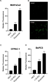
Fig. 6
Fig. 6
Compound 1-(S,Rp) induced DNA-single-strand breaks in recently replicated genomic DNA. (A) Concentration-dependent increase in single-stranded DNA breaks following treatment with 1-(S,Rp) (0–5 μM, 24 h) as assessed by the comet assay. (B) Pulse labelling with EdU (green fluorescence) prior to treatment confirms that DNA-strand breaks occur in recently replicated DNA following treatment with 1-(S,Rp) (5 μM, 24 h) but not when treated with NQO a direct acting genotoxic chemical, where only non-EdU labelled DNA counterstained with Hoechst (blue fluorescence) is visible in the comet tail. The results represent the mean of three independent biological experiments (±SD, n = 3), *** significantly different from untreated control (P < 0.001, 1-way ANOVA followed by a post hoc Dunnett's t-test).
Fig. 7
Fig. 7
Compound 1-(S,Rp) (10 μM, 24 h) induces double-stranded DNA breaks as assessed by gamma-H2AX phosphorylation in pancreatic ductal adeno carcinoma cells. (A) and (B) MIAPaCa2 cells as assessed by flow cytometry and confocal microscopy, respectively. (C) CFPAC-1 and (D) BxPC3 cells as assessed by flow cytometry. Etoposide (5 μM, 24 h) was used as a positive control. The results represent the mean of three independent biological experiments (±SD, n = 3). * and *** Statistically significantly different from control, P < 0.05 and 0.001, respectively (1-way ANOVA followed by a post hoc Dunnett's t-test).
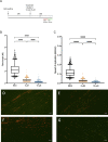
Fig. 8
Fig. 8
Compound 1-(S,Rp) inhibits DNA replication as assessed by single molecule DNA-fibre fluorography analysis. (A) Experimental design: Cells were seeded at a density of 3 × 105 cells/mL and allowed to attach for 24 h before treatment with 1-(S,Rp) (5, 10 and 25 μM) for 24 h before labelling with CldU (red) and IdU (green). (B) Total fibre length (red + green label) in kB and (C) Replication rate Kb/min are inhibited in a concentration-dependent manner by 1-(S,Rp). Box whisker plots show the mean (horizontal bar), interquartile range (box) and 5 and 95 percentile range (whisker) with values outside that range plotted as individual points. The results represent the mean of three independent experiments (n = 3) with 1072, 1406 and 1189 individual DNA fibres analysed in control, 5 and 10 μM treatments, respectively. Treatment with 25 μM 1-(S,Rp) resulted in complete inhibition of DNA fibres that were not quantified. *** Statistically significant (P < 0.0001) as assessed by a 1-way ANOVA followed by a post hoc Dunnett's t-test. Representative images from (D) Control, (E) 5 μM, (F) 10 μM and (G) 25 μM 1-(S,Rp) treated cells. Scale bar 10 μm and fibre length in micrometres was converted to kB of DNA as described in the materials and methods.
Fig. 9
Fig. 9
Compound 1-(S,Rp) stalls DNA replication in pancreatic ductal adenoma carcinoma cells resulting in activation of downstream signalling pathways and replication fork arrest. Highlighted in green are genes of this pathway that are statistically significantly transcriptionally activated in response to treatment with 1-(S,Rp). These include three out of the four genes involved in formation of the ‘9-1-1 checkpoint complex clamp’ (HUS1, Rad1 and Rad17) and two out of the three genes that form the MRN complex (MRE11 and NBN). Also detected experimentally and shown as red circles was phosphorylation of RPA1 and both checkpoint kinases 1 and 2. Diagram created in Biorender, www.biorender.com.

References

    1. Allemani C., Matsuda T., Di Carlo V., Harewood R., Matz M., Niksic M., Bonaventure A., Valkov M., Johnson C. J., Esteve J., Ogunbiyi O. J., Azevedo E. S. G., Chen W. Q., Eser S., Engholm G., Stiller C. A., Monnereau A., Woods R. R., Visser O., Lim G. H., Aitken J., Weir H. K., Coleman M. P., C W. Group, Global surveillance of trends in cancer survival 2000-14 (CONCORD-3): analysis of individual records for 37 513 025 patients diagnosed with one of 18 cancers from 322 population-based registries in 71 countries, The Lancet, 2018, 391 (10125), 1023–1075. - PMC - PubMed
    1. Conroy T., Desseigne F., Ychou M., Bouche O., Guimbaud R., Becouarn Y., Adenis A., Raoul J. L., Gourgou-Bourgade S., de la Fouchardiere C., Bennouna J., Bachet J. B., Khemissa-Akouz F., Pere-Verge D., Delbaldo C., Assenat E., Chauffert B., Michel P., Montoto-Grillot C., Ducreux M., Unicanc G. T. D., Intergrp P., FOLFIRINOX versus gemcitabine for metastatic pancreatic cancer, N. Engl. J. Med., 2011, 364 (19), 1817–1825. - PubMed
    1. Pacheco-Barcia V., France T., Zogopoulos G., Bouganim N., Donnay O., Alcindor T., Solis R. M., Guo K., Martin E., Colomer R., Asselah J., Gemcitabine plus nab-paclitaxel versus modified FOLFIRINOX as first line chemotherapy in metastatic pancreatic cancer: a comparison of toxicity and survival, Ann. Oncol., 2018, 29 (Suppl 5), v46.
    1. Williet N., Saint A., Pointet A. L., Tougeron D., Pernot S., Pozet A., Bechade D., Trouilloud I., Lourenco N., Hautefeuille V., Locher C., Desrame J., Artru P., Thirot Bidault A., Le Roy B., Pezet D., Phelip J. M., Taieb J., Folfirinox versus Gemcitabine/Nab-Paclitaxel as first-line therapy in patients with metastatic pancreatic cancer: a comparative propensity score study, Therap. Adv. Gastroenterol., 2019, 12, 175628481987866. - PMC - PubMed
    1. Cho I., Kang H., Jo J., Lee H., Chung M., Park J., Park S., Song S., Park M., An C., Jung S., Bang S., FOLFIRINOX versus gemcitabine plus Nab-Paclitaxel for treatment of metastatic pancreatic cancer: a single-center cohort study, Ann. Oncol., 2018, 29 (Suppl 5), v45.

Publication types