

T-cell proliferation assay for the detection of SARS-CoV-2-specific T-cells
- PMID: 35690083
- PMCID: PMC9174102
- DOI: 10.1016/j.cca.2022.05.025
T-cell proliferation assay for the detection of SARS-CoV-2-specific T-cells
Abstract
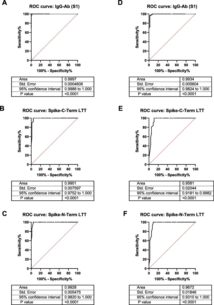
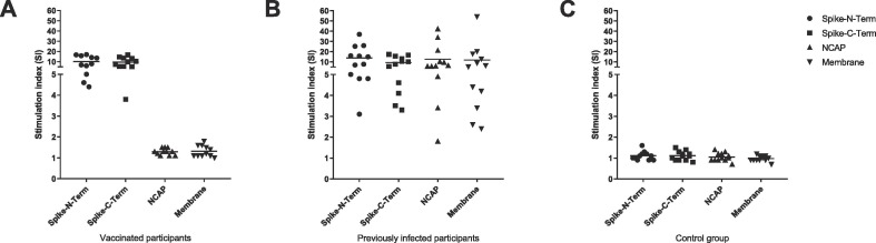
Both infection with and vaccination against SARS-CoV-2 trigger a complex B-cell and T-cell response. Methods for the analysis of the B-cell response are now well established. However, reliable methods for measuring the T-cell response are less well established and their usefulness in clinical settings still needs to be proven. Here, we have developed and validated a T-cell proliferation assay based on 3H thymidine incorporation. The assay is using SARS-CoV-2 derived peptide pools that cover the spike (S), the nucleocapsid (N) and the membrane (M) protein for stimulation. We have compared this novel SARS-CoV-2 lymphocyte transformation test (SARS-CoV-2 LTT) to an established ELISA assay detecting Immunoglobulin G (IgG) antibodies to the S1 subunit of the SARS-CoV-2 spike protein. The study was carried out using blood samples from both vaccinated and infected health care workers as well as from a non-infected control group. Our novel SARS-CoV-2 LTT shows excellent discrimination of infected and/or vaccinated individuals versus unexposed controls, with the ROC analysis showing an area under the curve (AUC) of > 0.95. No false positives were recorded as all unexposed controls had a negative LTT result. When using peptide pools not only representing the S protein (found in all currently approved vaccines) but also the N and M proteins (not contained in the vast majority of vaccines), the novel SARS-CoV-2 LTT can also discriminate T-cell responses resulting from vaccination against those induced by infection.
Keywords: Humoral and cellular immune responses; Lymphocyte transformation test; SARS-CoV-2; T-cell response.
Copyright © 2022 Elsevier B.V. All rights reserved.
Conflict of interest statement
The authors declare that they have no known competing financial interests or personal relationships that could have appeared to influence the work reported in this paper.
Figures
References
-
- Mahajan S., Kode V., Bhojak K., Karunakaran C., Lee K., Manoharan M., Ramesh A., Hv S., Srivastava A., Sathian R., Khan T., Kumar P., Gupta R., Chakraborty P., Chaudhuri A. Immunodominant T-cell epitopes from the SARS-CoV-2 spike antigen reveal robust pre-existing T-cell immunity in unexposed individuals. Sci. Rep. 2021;11(1) doi: 10.1038/s41598-021-92521-4. - DOI - PMC - PubMed
-
- Winkler E.S., Gilchuk P., Yu J., Bailey A.L., Chen R.E., Chong Z., Zost S.J., Jang H., Huang Y., Allen J.D., Case J.B., Sutton R.E., Carnahan R.H., Darling T.L., Boon A.C.M., Mack M., Head R.D., Ross T.M., Crowe J.E., Diamond M.S. Human neutralizing antibodies against SARS-CoV-2 require intact Fc effector functions for optimal therapeutic protection. Cell. 2021;184(7):1804–1820.e16. - PMC - PubMed
-
- Zuo J., Dowell A.C., Pearce H., Verma K., Long H.M., Begum J., Aiano F., Amin-Chowdhury Z., Hoschler K., Brooks T., Taylor S., Hewson J., Hallis B., Stapley L., Borrow R., Linley E., Ahmad S., Parker B., Horsley A., Amirthalingam G., Brown K., Ramsay M.E., Ladhani S., Moss P. Robust SARS-CoV-2-specific T cell immunity is maintained at 6 months following primary infection. Nat. Immunol. 2021;22(5):620–626. - PMC - PubMed
MeSH terms
Substances
LinkOut - more resources
Full Text Sources
Medical
Miscellaneous
