

Progesterone and its receptor signaling in cervical remodeling: Mechanisms of physiological actions and therapeutic implications
- PMID: 35690241
- PMCID: PMC9509468
- DOI: 10.1016/j.jsbmb.2022.106137
Progesterone and its receptor signaling in cervical remodeling: Mechanisms of physiological actions and therapeutic implications
Abstract
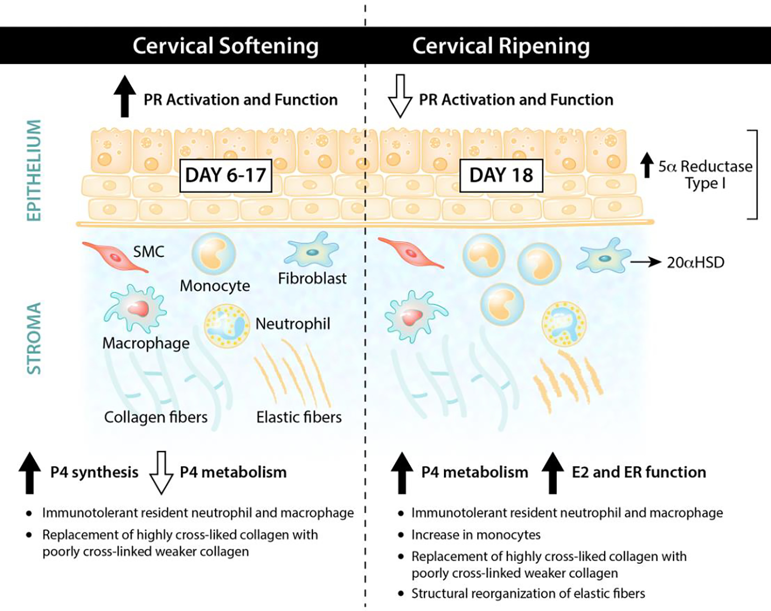
The remodeling of the cervix from a closed rigid structure to one that can open sufficiently for passage of a term infant is achieved by a complex series of molecular events that in large part are regulated by the steroid hormones progesterone and estrogen. Among hormonal influences, progesterone exerts a dominant role for most of pregnancy to initiate a loss of tissue strength yet maintain competence in a phase termed softening. Equally important are the molecular events that abrogate progesterone function in late pregnancy to allow a loss of tissue competence and strength during cervical ripening and dilation. In this review, we focus on current understanding by which progesterone receptor signaling for the majority of pregnancy followed by a loss/shift in progesterone receptor action at the end of pregnancy, collectively ensure cervical remodeling as necessary for successful parturition.
Keywords: Cervical remodeling; Cervix and cervical ripening; Pregnancy; Preterm birth; Progesterone; Progesterone receptor.
Copyright © 2022 Elsevier Ltd. All rights reserved.
Figures
Similar articles
-
Dynamics of cervical remodeling during pregnancy and parturition: mechanisms and current concepts.Semin Reprod Med. 2007 Jan;25(1):69-79. doi: 10.1055/s-2006-956777. Semin Reprod Med. 2007. PMID: 17205425 Review.
-
Contractile function of the cervix plays a role in normal and pathological pregnancy and parturition.Med Hypotheses. 2020 Dec;145:110336. doi: 10.1016/j.mehy.2020.110336. Epub 2020 Oct 7. Med Hypotheses. 2020. PMID: 33049595 Free PMC article.
-
Estrogen and progesterone metabolism in the cervix during pregnancy and parturition.J Clin Endocrinol Metab. 2008 Jun;93(6):2366-74. doi: 10.1210/jc.2007-2813. Epub 2008 Mar 25. J Clin Endocrinol Metab. 2008. PMID: 18364378 Free PMC article.
-
Cervix Stromal Cells and the Progesterone Receptor A Isoform Mediate Effects of Progesterone for Prepartum Remodeling.Reprod Sci. 2019 May;26(5):690-696. doi: 10.1177/1933719118820462. Epub 2019 Jan 17. Reprod Sci. 2019. PMID: 30654718 Free PMC article.
-
Contributions to the dynamics of cervix remodeling prior to term and preterm birth.Biol Reprod. 2017 Jan 1;96(1):13-23. doi: 10.1095/biolreprod.116.142844. Biol Reprod. 2017. PMID: 28395330 Free PMC article. Review.
Cited by
-
Pregnancy influences expression of interferon-stimulated genes, progesterone receptor and progesterone-induced blocking factor in ovine thyroid.Anim Biosci. 2024 Aug;37(8):1377-1386. doi: 10.5713/ab.23.0508. Epub 2024 Apr 25. Anim Biosci. 2024. PMID: 38665088 Free PMC article.
-
Effects of dietary genistin supplementation on reproductive performance, immunity and antioxidative capacity in gestating sows.Front Vet Sci. 2024 Nov 21;11:1489227. doi: 10.3389/fvets.2024.1489227. eCollection 2024. Front Vet Sci. 2024. PMID: 39641093 Free PMC article.
-
Promegestone Prevents Lipopolysaccharide-Induced Cervical Remodeling in Pregnant Mice.Cells. 2025 Feb 7;14(4):242. doi: 10.3390/cells14040242. Cells. 2025. PMID: 39996716 Free PMC article.
-
In Vitro and Ex Vivo Evaluation of Novel Methacrylated Chitosan-PNIPAAm-Hyaluronic Acid Hydrogels Loaded with Progesterone for Applications in Vaginal Delivery.Polymers (Basel). 2024 Jul 30;16(15):2160. doi: 10.3390/polym16152160. Polymers (Basel). 2024. PMID: 39125186 Free PMC article.
-
Preterm Birth and Corticotrophin-Releasing Hormone as a Placental Clock.Endocrinology. 2022 Dec 19;164(2):bqac206. doi: 10.1210/endocr/bqac206. Endocrinology. 2022. PMID: 36478045 Free PMC article. Review.
References
-
- Yoshida K, Jiang H, Kim M, Vink J, Cremers S, Paik D, Wapner R, Mahendroo M, Myers K, Quantitative evaluation of collagen crosslinks and corresponding tensile mechanical properties in mouse cervical tissue during normal pregnancy, PLoS One, 9 (2014) e112391. 10.1371/journal.pone.0112391. - DOI - PMC - PubMed
Publication types
MeSH terms
Substances
Grants and funding
LinkOut - more resources
Full Text Sources
Research Materials
