. 2022 Jun 11;13(1):3372.
doi: 10.1038/s41467-022-31109-6.

Mechanism of glycogen synthase inactivation and interaction with glycogenin

Affiliations

Mechanism of glycogen synthase inactivation and interaction with glycogenin

Laura Marr et al. Nat Commun. .

Abstract

Glycogen is the major glucose reserve in eukaryotes, and defects in glycogen metabolism and structure lead to disease. Glycogenesis involves interaction of glycogenin (GN) with glycogen synthase (GS), where GS is activated by glucose-6-phosphate (G6P) and inactivated by phosphorylation. We describe the 2.6 Å resolution cryo-EM structure of phosphorylated human GS revealing an autoinhibited GS tetramer flanked by two GN dimers. Phosphorylated N- and C-termini from two GS protomers converge near the G6P-binding pocket and buttress against GS regulatory helices. This keeps GS in an inactive conformation mediated by phospho-Ser641 interactions with a composite "arginine cradle". Structure-guided mutagenesis perturbing interactions with phosphorylated tails led to increased basal/unstimulated GS activity. We propose that multivalent phosphorylation supports GS autoinhibition through interactions from a dynamic "spike" region, allowing a tuneable rheostat for regulating GS activity. This work therefore provides insights into glycogen synthesis regulation and facilitates studies of glycogen-related diseases.


Conflict of interest statement

The authors declare no competing interests.

Figures

Fig. 1
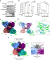
Fig. 1. Structural analysis of the full-length GS-GN complex.
a Enzymatic reaction catalysed by GN. b Enzymatic reaction catalysed by GS and subsequent branching of glycogen by GBE. c Domain architecture of human GS (top) and GN (bottom). Known in vivo phosphorylation sites of GS are shown in red and are labelled with residue number and classical nomenclature (in bold). GN tyrosine 195 that becomes auto-glucosylated and was mutated to a phenylalanine (Y195F) in this study is indicated. Not to scale. d SDS-PAGE analysis of GS-GN WT and Y195F complexes (left) and periodic acid-Schiff (PAS) staining of both complexes (right). Source data are provided as a Source Data file. Data are representative of three independent experiments. e Cartoon representation of GN WT and Y195F. f Mass photometry of GS-GN(Y195F) (left) and WT complex (right). Expected stoichiometry for each peak is indicated. The percentage of particles contributing to each peak is shown in brackets. g Selected 2D class averages after negative-stain electron microscopy (nsEM) analysis of indicated GS-GN complexes. Data are representative of 84,420 and 4419 particles for the GS-GN(Y195F) and GS-GN complex respectively. h Negative stain EM final map (C1 symmetry at ~22 Å) is shown in transparent surface, with fitted human GN crystal structure (PDB ID 3T7O) and human GS cryo-EM structure (reported here).
Fig. 2
Fig. 2. Cryo-EM structure of human GS-GN34 complex.
a Immunoblot for the indicated human GS phosphorylation sites and total GS. Data are representative of two independent experiments carried out in technical duplicates. b Activity of GS-GN(Y195F) with and without the addition of lambda protein phosphatase (lambda PP) and protein phosphatase 1 (PP1) (left) and −/+ G6P activity ratio (right). Upon G6P saturation, GS reaches similar activity levels regardless of phosphorylation state. Data are mean ± S.E.M. from n = 2 and representative of two independent experiments. One-way analysis of variance (Tukey’s post hoc test); exact p values are shown. Source data for (a and b) are provided as a Source Data file. c 2.6 Å cryo-EM map of the GS tetramer coloured by corresponding chain. Density corresponding to the GN34 C-terminal region is shown in green. d Human GS-GN34 cartoon model shown in ribbons coloured by corresponding chain (left). Interaction between GS and GN34 (right). e Unsharpened cryo-EM map shown at a lower threshold to visualise the “spike” region depicted in grey (left). The N- and C-terminal tails of two protomers converge and form the “spike” region (right).
Fig. 3
Fig. 3. The phosphoregulatory region of human GS.
a Human (Hs)GS-GN34 structure shown in ribbons (top left). The N- and C- terminal tails of one GS protomer (chain A) lie next to one another and move towards the adjacent protomer, meeting the N- and C-terminal tails from chain B. Arrows indicate continuation of cryo-EM density (top right). Electron density (C1 symmetry) for phosphorylated S641 (pS641) interacting with R588 and R591 on the regulatory helices α22 (bottom left). Residues that are interacting with the N- and C-terminal tails that are mutated in this study are shown (bottom right). b Comparison of distances between regulatory helices of adjacent monomers of HsGS (reported here), low activity inhibited mimic (PDB ID 5SUL), basal state (PDB ID 3NAZ) and G6P activated (PDB ID 5SUK) yeast GS (yGS) crystal structures. Quoted distances were measured from Cα of Arg591 (chain A) and -Cα of Arg580 (chain B) of HsGS and corresponding yeast residues.
Fig. 4
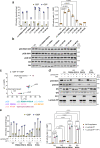
Fig. 4. Dislodging the GS phosphoregulatory region increases basal activity and increases accessibility for phosphatases.
a Activity of GS WT and indicated mutants in the GS-GN(Y195F) complex in the presence and absence of glucose-6-phopshate (G6P) (left) and −/+ G6P activity ratio (right). Data are mean ± S.E.M from n = 3 and representative of two independent experiments. One-way analysis of variance, (Tukey’s post hoc test); exact p values are shown. b Western blot for human GS phosphorylation sites S641/645, S641, S8, and total GS and GN. Data are representative of two independent experiments carried out in technical duplicates. c Melting temperature (Tm) of GS WT and mutants in the GS-GN(Y195F) complex. Changes in melting temperature upon addition of 12.5 mM G6P (ΔTm = Tm+G6P–Tm–G6P). Data are mean ± S.E.M from n = 3 experiments carried out in technical duplicates (dephosphorylated GS) and triplicates (WT and mutant GS). d Western blots of GS WT and mutants in the GS-GN(Y195F) complex after dephosphorylation with protein phosphatase 1 (PP1) and/or lambda protein phosphatase (lambda PP). Data are representative of three independent experiments carried out in technical duplicates. e Activity of phosphorylated and dephosphorylated GS WT and indicated mutants (left) and −/+ G6P activity ratio (right). Data are mean ± S.E.M from n = 2 and representative of two independent experiments. Two-way analysis of variance (Tukey’s post hoc test); exact p values are shown. Source data for all panels are provided as a Source Data file.
Fig. 5
Fig. 5. GS and GN cooperate to synthesise glycogen.
Glucose is converted into glycogen through the action of glycogenin (GN), glycogen synthase (GS) and glycogen branching enzyme (GBE). GN interacts with GS to feed the initial glucose chain into the GS active site for elongation. GS is regulated by allosteric activation and inhibitory phosphorylation. Phospho-S641 (pS641) from one C- terminal tail interacts with the regulatory helices α22 to cause enzyme inhibition. This can be relieved by glucose-6-phoshate (G6P), with or without phosphatases, to reach a high activity state. Kinases can phosphorylate GS to inhibit the enzyme.

References

    1. Roach PJ, Depaoli-Roach AA, Hurley TD, Tagliabracci VS. Glycogen and its metabolism: some new developments and old themes. Biochem. J. 2012;441:763–787. doi: 10.1042/BJ20111416. - DOI - PMC - PubMed
    1. Roach PJ. Glycogen and its metabolism. Curr. Mol. Med. 2002;2:101–120. doi: 10.2174/1566524024605761. - DOI - PubMed
    1. Pitcher J, Smythe C, Campbell DG, Cohen P. Identification of the 38‐kDa subunit of rabbit skeletal muscle glycogen synthase as glycogenin. Eur. J. Biochem. 1987;169:497–502. doi: 10.1111/j.1432-1033.1987.tb13637.x. - DOI - PubMed
    1. Zeqiraj E, Sicheri F. Getting a handle on glycogen synthase – Its interaction with glycogenin. Mol. Asp. Med. 2015;46:63–69. doi: 10.1016/j.mam.2015.08.004. - DOI - PMC - PubMed
    1. Skurat AV, Dietrich AD, Roach PJ. Interaction between glycogenin and glycogen synthase. Arch. Biochem. Biophys. 2006;456:93–97. doi: 10.1016/j.abb.2006.09.024. - DOI - PMC - PubMed

Publication types

MeSH terms