. 2022 Jun 11;12(1):9674.
doi: 10.1038/s41598-022-13405-9.

Muscle regeneration affects Adeno Associated Virus 1 mediated transgene transcription

Affiliations

Muscle regeneration affects Adeno Associated Virus 1 mediated transgene transcription

Amédée Mollard et al. Sci Rep. .

Abstract

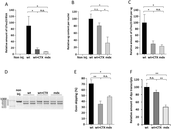
Duchenne muscular dystrophy is a severe neuromuscular disease causing a progressive muscle wasting due to mutations in the DMD gene that lead to the absence of dystrophin protein. Adeno-associated virus (AAV)-based therapies aiming to restore dystrophin in muscles, by either exon skipping or microdystrophin expression, are very promising. However, the absence of dystrophin induces cellular perturbations that hinder AAV therapy efficiency. We focused here on the impact of the necrosis-regeneration process leading to nuclear centralization in myofiber, a common feature of human myopathies, on AAV transduction efficiency. We generated centronucleated myofibers by cardiotoxin injection in wild-type muscles prior to AAV injection. Intramuscular injections of AAV1 vectors show that transgene expression was drastically reduced in regenerated muscles, even when the AAV injection occurred 10 months post-regeneration. We show also that AAV genomes were not lost from cardiotoxin regenerated muscle and were properly localised in the myofiber nuclei but were less transcribed leading to muscle transduction defect. A similar defect was observed in muscles of the DMD mouse model mdx. Therefore, the regeneration process per se could participate to the AAV-mediated transduction defect observed in dystrophic muscles which may limit AAV-based therapies.


Conflict of interest statement

The authors declare no competing interests.

Figures

Figure 1
Figure 1
Description of cardiotoxin regenerated and mdx muscles. Tibialis anterior (TA) muscles of wild-type (wt) mice were injected or not with 0.5 nmol of cardiotoxin (CTX) to induce muscle regeneration. Six weeks after injury TAs were collected and analysed. (A) Number of myofibers per mm2 of wt, wt + CTX and mdx muscles. (B) Quantification of cross-sectional area (CSA) mean. (C) Muscle mass. (D) Distribution of myofiber CSA. (E) Percentage of fibers expressing I, IIa, IIx and IIb myosin heavy chain isoforms. (F) Counts of muscle nuclei classified into interstitial, peripheral and central localization. The data represent the mean values of four TAs per group ± SEM. n.s., non-significant, *P ≤ 0.05, **P ≤ 0.01, ***P ≤ 0.001. For D, *P ≤ 0.05 between wt + CTX group and wt or mdx groups, + P ≤ 0.05 between wt group and wt + CTX or mdx groups.
Figure 2
Figure 2
Evaluation of AAV1-mSeAP expression in cardiotoxin regenerated and mdx muscles. (A) TA muscles of wt mice were injected or not with 0.5 nmol of cardiotoxin (CTX) to induce muscle regeneration. Three weeks or 42 weeks later, the CTX muscles (wt + CTX), as well as wt muscles (wt) were injected with AAV1-mSeAP vector. Muscles were analysed 3 weeks after vector injection. TA muscles of mdx mice were also injected with AAV1-mSeAP vector and analysed 3 weeks after injection. Wt TAs analysed at 6 weeks = 5, wt + CTX at 6 weeks = 5, mdx = 6, wt at 45 weeks = 5, wt + CTX at 45 weeks = 5. (B) Representative (top) haematoxylin and eosin (HE) staining and (bottom) histochemical detections of mSeAP activity in TA transversal sections of non-injected wt (non inj.), wt, wt + CTX and mdx muscles 6 weeks after CTX injury and 3 weeks after AAV1-mSeAP injection. The quantification of the centronucleated fibers expressed as percent of total fibers as well as mSeAP activity is shown. (C) Representative haematoxylin and eosin (HE) staining and histochemical detections of mSeAP activity in TA transversal sections of wt and wt + CTX muscles 45 weeks after CTX injury and 3 weeks after AAV1-mSeAP injection. The quantification of the centronucleated fibers expressed as percent of total fibers as well as mSeAP activity is shown. ***p < 0.001, ****p < 0.0001. n.s., non-significant. Scale bar 100 µm.
Figure 3
Figure 3
Impact of muscle regeneration on vector genome content and its intracellular localization. (A) Quantification of AAV genomes per nucleus by absolute qPCR in wt, wt + CTX and mdx muscles injected with AAV1-mSeAP vector. Three weeks after AAV injections, TAs were collected and analysed. Considering the higher numbers of total nuclei in wt + CTX and mdx muscles compared to wt muscles, the obtained data for these muscles were adjusted by a correcting factor (1.7 and 1.5, respectively). AAV genome content is expressed as the AAV genome number relative to the value obtained for wt muscles. Non inj., wt non-AAV injected TA. (B) Western blot analysis of total (T), nuclear (N) and cytosolic (C) fractions from the three muscle groups using GAPDH and H3 antibodies, confirming cytosolic and nuclear enrichments. EEA1 and GM130 antibodies detect markers of early endosomes and Golgi apparatus present in the cytosolic fractions validating the nuclear fraction purity. (D) AAV genomes were quantified by absolute qPCR in nuclear and total fractions from the three muscle groups and the proportion of vg present in nuclear fractions was calculated. (D) Quantification of AAV genomes per nucleus by absolute qPCR 10 month after regeneration. Considering the higher numbers of total nuclei in wt + CTX muscles compared to wt muscles, the obtained data for wt + CTX were adjusted by the correcting factor 1.7. AAV genome content is expressed as the AAV genome number relative to the value obtained for wt muscles (6w). Non inj., wt non-AAV injected TA. The data represent the mean values of five TAs per group ± SEM. n.s., non-significant, ***P ≤ 0.001.
Figure 4
Figure 4
Evaluation of AAV1-mSeAP expression after muscle regeneration. (A) Quantification of mSeAP pre-messengers (pre-mRNA) and transcripts (mRNA) 6 weeks after CTX injury and 3 week after AAV-mSeAP injection performed by relative qPCR and normalised by the AAV genome numbers. Relative RNA number is expressed as a percentage of wt RNAs. (B) Quantification of mSeAP mRNA at 6 and 45 weeks post-injury by relative qPCR and expressed as a percentage of mSeAP transcripts present in wt muscles (6w). Non inj., wt non-AAV injected TA. (C) Quantification of mSeAP transcripts normalised by the AAV genome numbers in the corresponding muscles. Relative RNA number is expressed as a percentage of wt transcripts (6w). The data represent the mean values of minimum four TAs per group ± SEM. N.s. non-significant, *P ≤ 0.05, **P ≤ 0.01, ***P ≤ 0.001.
Figure 5
Figure 5
Evaluation of AAV1-U7ex23 benefit in cardiotoxin regenerated and mdx muscles. Wt, wt + CTX and mdx TAs were injected with AAV1-U7ex23 and the mice were sacrificed 3 weeks later. (A) Level of U7ex23 RNA estimated by relative qPCR and expressed as a percentage of U7ex23 present in wt muscles. Non inj., wt non-AAV injected TA. (B) Quantification of AAV genomes per nucleus by absolute qPCR in wt, wt + CTX and mdx muscles injected with AAV- U7ex23 vector and corrected by the correcting factors. AAV genome content is expressed as the AAV genome number relative to the value obtained for wt muscles. (C) U7ex23 RNA amount normalised by the AAV genome numbers and expressed as a percentage of wt RNAs. (D) Level of exon 23 skipping estimated by nested RT-PCR. The 901 bp PCR product corresponds to full-length dystrophin transcripts whereas the 688 bp product corresponds to transcripts lacking exon 23. (E) Quantification of exon 23 skipping by relative qPCR and expressed as a percentage of total dystrophin transcripts. (F) Quantification of dystrophin (dys) transcripts by relative qPCR. Relative amount of dys transcripts is expressed as a percentage of wt RNAs. The data represent the mean values of four TAs per group ± SEM. Student’s t-test: n.s. non-significant, *P ≤ 0.05, **P ≤ 0.01.

Similar articles

Cited by

References

    1. Davies KE, Nowak KJ. Molecular mechanisms of muscular dystrophies: old and new players. Nat. Rev. Mol. Cell Biol. 2006;7:762–773. doi: 10.1038/nrm2024. - DOI - PubMed
    1. Duan D, Goemans N, Takeda S, Mercuri E, Aartsma-Rus A. Duchenne muscular dystrophy. Nat. Rev. Dis. Primers. 2021;7:13. doi: 10.1038/s41572-021-00248-3. - DOI - PMC - PubMed
    1. Duguez S, et al. Dystrophin deficiency leads to disturbance of LAMP1-vesicle-associated protein secretion. Cell Mol. Life Sci. 2013;70:2159–2174. doi: 10.1007/s00018-012-1248-2. - DOI - PMC - PubMed
    1. Le Guiner C, et al. Forelimb treatment in a large cohort of dystrophic dogs supports delivery of a recombinant AAV for exon skipping in Duchenne patients. Mol. Ther. 2014;22:1923–1935. doi: 10.1038/mt.2014.151. - DOI - PMC - PubMed
    1. Le Guiner C, et al. Long-term microdystrophin gene therapy is effective in a canine model of Duchenne muscular dystrophy. Nat. Commun. 2017;8:16105. doi: 10.1038/ncomms16105. - DOI - PMC - PubMed

Publication types

Supplementary concepts