. 2022 Jun 11;12(1):9693.
doi: 10.1038/s41598-022-13807-9.

Mechanical loading of cranial joints minimizes the craniofacial phenotype in Crouzon syndrome

Affiliations

Mechanical loading of cranial joints minimizes the craniofacial phenotype in Crouzon syndrome

Mehran Moazen et al. Sci Rep. .

Abstract

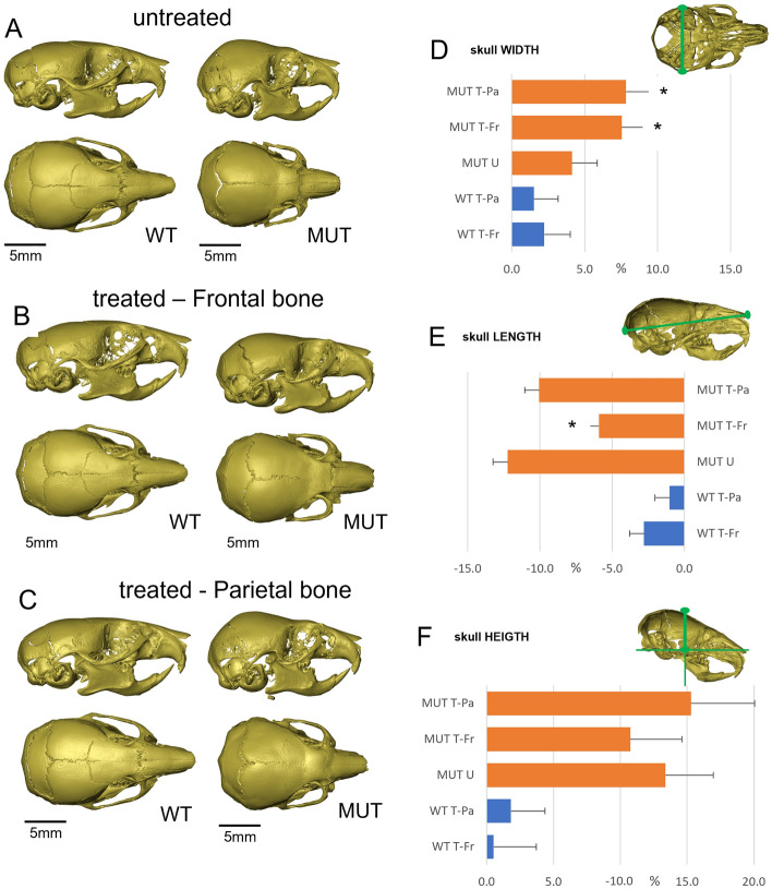
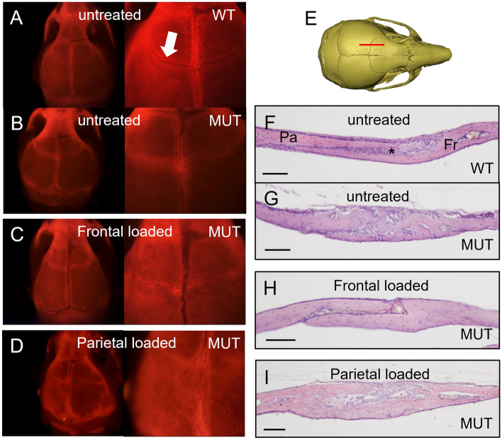
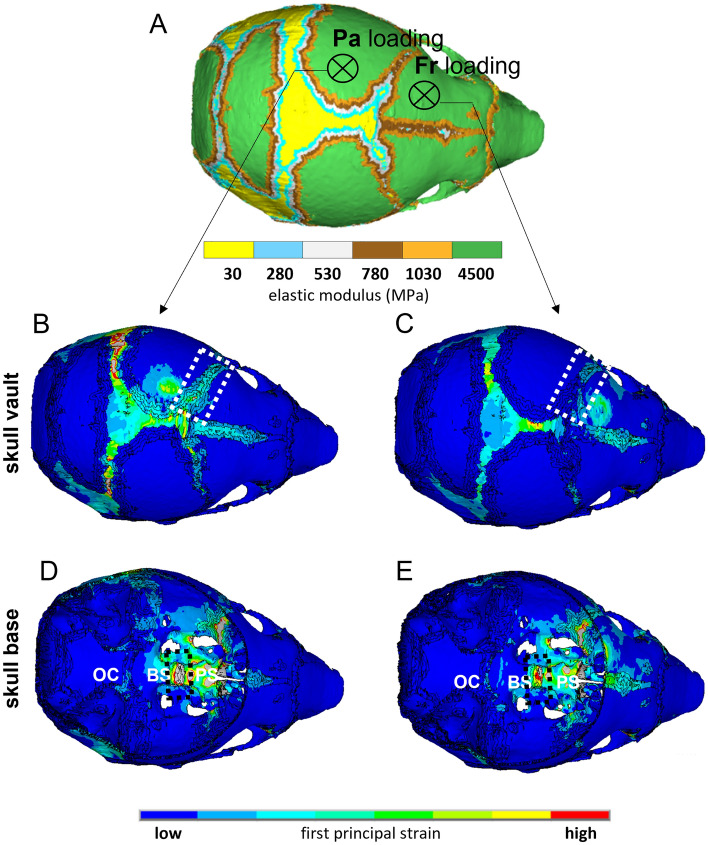
Children with syndromic forms of craniosynostosis undergo a plethora of surgical interventions to resolve the clinical features caused by the premature fusion of cranial sutures. While surgical correction is reliable, the need for repeated rounds of invasive treatment puts a heavy burden on the child and their family. This study explores a non-surgical alternative using mechanical loading of the cranial joints to prevent or delay craniofacial phenotypes associated with Crouzon syndrome. We treated Crouzon syndrome mice before the onset of craniosynostosis by cyclical mechanical loading of cranial joints using a custom designed set-up. Cranial loading applied to the frontal bone partially restores normal skull morphology, significantly reducing the typical brachycephalic appearance. This is underpinned by the delayed closure of the coronal suture and of the intersphenoidal synchondrosis. This study provides a novel treatment alternative for syndromic craniosynostosis which has the potential to be an important step towards replacing, reducing or refining the surgical treatment of all craniosynostosis patients.


Conflict of interest statement

The authors declare no competing interests.

Figures

Figure 1
Figure 1
Cranial bone loading on pre-weaning Crouzon mice using actuator technology. (A) Equipment set-up showing P10 mouse in a custom-designed, 3D-printed anaesthetic mask and holder, with a window for access to the skull vault. The electrical, linear actuator moves a small probe (1 mm) that loads the bone underneath, a sensor records the force applied. (B,C) Close-ups of the loading protocol in progress at postnatal day 7 (B) and 21 (C) with the actuator probe targeting the frontal bone. (D) Top view of a micro-CT generated image of a wild-type mouse skull (P21). The asterisks indicate the approximate loading locations on the parietal (Pa) and frontal (Fr) bones, either side of the coronal suture. (E) Histological, sagittal section (H&E) through an adult mouse skull showing the parietal (Pa) and frontal (Fr) bones and the suture mesenchyme (SM) situated between the overlapping parts of the parietal and frontal bones. (F) Schematic representation of the mechanical action generated by loading on either the parietal (Pa) or frontal (Fr) bones. Loading the frontal bone will generate tensile forces on the suture (right) and loading the parietal bone will generate compression forces on the suture (left). (G) Treatment schedule for the cranial loading protocol starting at P7 and loading 10 days until P18. Skulls are collected at P21.
Figure 2
Figure 2
Changes in Crouzon skull morphology after cranial bone loading. (A) Micro-CT images of untreated P21 skulls. MUT (Fgfr2_C342Y) skulls show typical signs of brachycephaly and synostosis of the coronal suture. (B) Micro-CT images P21 skulls loaded on the frontal bone. While WT skulls appear unchanged, MUT skulls display a partially restored skull shape with a reduced brachycephalic appearance. Synostosis of the coronal suture does not appear to be affected. (C) Micro-CT images P21 skulls loaded on the parietal bone. Compared to untreated skulls, no changes can be observed. (D–F) Graphs representing relative skull dimensions in percentage increase/decrease compared to untreated WT controls. (D) Skull measurements show that frontal and parietal loading increases mutant skull width by respectively 7.5% and 7.8% as compared to 4.1% in untreated mutant skulls. (E) Untreated, mutant skulls are 12.2% shorter than untreated WT skulls, indicating the midfacial hypoplasia phenotype. Only frontal bone loaded skulls show a statistically significant difference and are only 5.9% shorter. (F) Untreated, mutant skulls are 13.4% higher than untreated WT skulls, indicating the brachycephalic appearance of these skulls. No statistically significant differences are found for relative height in treated skulls. Images in (AC) are representative of the average dimensions per experimental group. Untreated-WT n = 12, frontal loaded-WT (WT T-Fr) n = 15, parietal loaded-WT (WT T-Pa) n = 9, untreated MUT (MUT U) n = 16, frontal loaded MUT (MUT T-Fr) n = 7, parietal loaded MUT (MUT T-Pa) n = 8. *Statistically significant (p < 0.05) based on t test performed against MUT U. Error bars in (DF) represent approximate variance calculated using the Delta method.
Figure 3
Figure 3
Changes in coronal suture morphology and histology following cranial bone loading. (A–D) Calvaria showing fluorescent Alizarin complexone staining at 0.8 × (left) and 2 × (right) magnification. (A) Wild-type (WT) calvaria show the typical overlapping bone structure of the coronal suture (arrow). (B) Mutant (MUT) coronal sutures show some staining indicating active bone growth but the overlapping structure is absent. (C) Frontal loaded calvaria show a patent suture, but with a reduced area of frontal-parietal overlap. (D) Parietal loaded calvaria show no difference from untreated mutant sutures. (E) Sagittal sections through the coronal suture (red line) at P21 were stained with H&E. (F) Wild-type (WT) sutures show a characteristic overlapping feature of the parietal (P) and frontal (F) bones with the suture mesenchyme (*) in between. (G) Mutant (MUT) sutures show a loss of patency phenotype with fusion between parietal and frontal bones and loss of mesenchyme. The suture appears thickened as compared to flanking bone. (H) Animals treated with frontal bone loading show a more normal suture with bones appearing unfused, but with significant loss of mesenchyme. (I) Animals treated with parietal bone loading show no improvement and look similar to untreated sutures. Scale bars are 100 µm.
Figure 4
Figure 4
Changes in skull base morphology and histology following cranial bone loading. (A) Schematic of the skull base showing anatomical structures. OC occipital, BS basi-sphenoid, PS pre-sphenoid, SOS spheno-occipital synchondrosis, ISS intersphenoidal synchondrosis. (BD) Micro-CT images of sagittal, midline sections through the skull. Untreated WT skulls show a fully patent ISS (inset) while MUT synchondroses have closed (B). Frontal loaded skulls show improved patency of the ISS (C), while parietal loaded skulls do not (D). (E) Patency of the anterior synchondrosis (ISS) was scored blinded for sample identity. While all WT synchondroses are open (green) at P21, the majority of the untreated MUT skull bases have a partially (yellow) or fully closed (red) ISS. Skulls loaded on the frontal bone show a statistically significant improvement while parietal loaded skulls do not. Untreated-WT (WT U) n = 12, frontal loaded-WT (WT T-Fr) n = 15, parietal loaded-WT (WT T-Pa) n = 9, untreated MUT (MUT U) n = 16, frontal loaded MUT (MUT T-Fr) n = 7, parietal loaded MUT (MUT T-Pa) n = 8. (F) Sagittal, midline sections through the skull base (red line) at P21 were stained with H&E. (G–J) Histological analysis of the Crouzon-associated phenotypic features following cranial loading. (G) A wild-type skull base showing the bones separated by synchondroses. SOS spheno-occipital synchondrosis, ISS intersphenoidal synchondrosis. The magnified image of the ISS shows a rectangular cartilage situated between endochondral bone. (H) Mutant (MUT) ISS shows an aberrant cone shape and the cartilage structure protrudes cranially. (I) Animals treated with frontal bone loading show a more normal synchondroses. (J) Animals treated with parietal bone loading show no improvement and look similar to untreated synchondroses.
Figure 5
Figure 5
Comparing the pattern of mechanical strain distribution across the skull between frontal and parietal loading. (A) Finite element model of a P3 mouse skull that was computationally grown to P7 and used to investigate the difference in the pattern of strain distribution due to the frontal and parietal loading. It indicates the mechanical properties of different skull regions and the anatomical position that were loaded analogous to the in vivo experiments. Heat maps were generated using ANSYS v.18 (ANSYS Inc., Canonsburg, PA, USA). (B,C) Comparing the pattern of first principal strain across the skull roof between parietal and frontal loading respectively. The dashed box highlights the coronal suture. (D,E) Comparing the pattern of first principal strain across the skull base between parietal and frontal loading respectively. The dashed box highlights the ISS (intersphenoidal synchondrosis). OC occipital, BS basi-sphenoid, PS pre-sphenoid.

Similar articles

Cited by

References

    1. Lajeunie E, Le Merrer M, Bonaiti-Pellie C, Marchac D, Renier D. Genetic study of nonsyndromic coronal craniosynostosis. Am. J. Med. Genet. 1995;55:500–504. doi: 10.1002/ajmg.1320550422. - DOI - PubMed
    1. EUROCAT. European Platform on Rare Disease Registration-Prevalence. https://eu-rd-platform.jrc.ec.europa.eu/eurocat/eurocat-data/prevalence_en (2021).
    1. Boyadjiev SA, International Craniosynostosis Consortium Genetic analysis of non-syndromic craniosynostosis. Orthod. Craniofac. Res. 2007;10:129–137. doi: 10.1111/j.1601-6343.2007.00393.x. - DOI - PubMed
    1. Cunningham ML, Seto ML, Ratisoontorn C, Heike CL, Hing AV. Syndromic craniosynostosis: From history to hydrogen bonds. Orthod. Craniofac. Res. 2007;10:67–81. doi: 10.1111/j.1601-6343.2007.00389.x. - DOI - PubMed
    1. Wilkie AO, et al. Prevalence and complications of single-gene and chromosomal disorders in craniosynostosis. Pediatrics. 2010;126:e391–e400. doi: 10.1542/peds.2009-3491. - DOI - PMC - PubMed

Publication types